
Zazubovits Research Group

Our research interests include:

  • Renewable energy: Optical spectroscopy of pigment-protein light-harvesting photosynthetic complexes - energy transfer and charge transfer;
  • Biophotonics / protein structure and [low-temperature] dynamics: Protein-chlorophyll complexes as model systems. Heat transfer in proteins and through protein-water interfaces. Energy landscapes and dynamics in amorphous solids;
  • Biosensors for explosives detection utilizing photosynthetic reaction centers;
  • Combining optical (Raman) and transport measurements in graphene and carbon nanotubes.
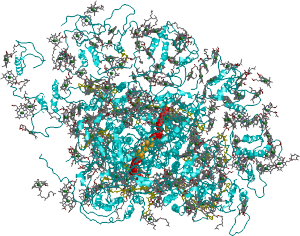
Top view of plant Photosystem I. Blue: protein. Grey: chlorophylls. Yellow: carotenoids. Red: quinones, Orange: FeS clusters.

The energy needs of the humankind are constantly increasing. Satisfying these needs by burning fossil fuels results in pollution, undesired climate changes and eventual exhaustion of the fuel reserves. Nuclear energy is not problem-free either. Renewable energy is likely to provide the solution for these challenges. The sun offers the most abundant potential source of energy and delivers 3,850,000x1018 J per year to Earth, compared to ~630 x1018 J per year currently consumed by the human civilization. Sun energy was supporting the life on Earth for billions of years via process of photosynthesis. (In fact, all existing fossil fuel reserves originate from photosynthesis processes of long ago.) As a result of evolution, proteins performing the first steps of photosynthesis achieved amazing effectiveness. Can we learn something useful from Nature by exploring how these first steps are organized? To this end, we explore energy and primary charge separation processes in isolated photosynthetic proteins.

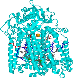
Side view of the bacterial reaction center (BRC). Grey: bacteriochlorophylls. Dark-blue: bacteriopheophytines, Red: quinones, Orange: non-heme iron. Nobel Prize in Chemistry was awarded for determining BRC structure in 1988

Proteins involved in photosynthesis can also serve as model systems in the studies of some very general protein features. Proteins perform wide variety of tasks in the living organisms. Their ability to perform the designated tasks depends critically on their tertiary structure, achieved as a result of folding. But how do proteins “know” how to fold properly? It is well known that they fold to minimize their free energy (including electrostatic and entropic terms). However, folding even a small protein by trial and error would take forever (Levinthal paradox). Thus, proteins are believed to possess funnel-like energy landscapes with multiple hierarchal tiers. These landscapes can be explored using optical spectroscopy. Proteins involved in photosynthesis have in-built fluorescent molecules (chlorophylls) that can serve as very sensitive probes to local protein environment. Optical spectroscopy also allows one to explore heat transport in proteins and through protein-water interfaces.

Photosynthetic reaction centers utilize light energy to transfer electrons through the (thylakoid or cell) membrane. A number of substances can inhibit this process. (The strongest of these substances are agricultural herbicides.) We have demonstrated inhibition of electron transfer in isolated photosynthetic reaction centers by explosives and are continuing to work on photosynthesis-based explosives biosensors.

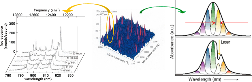
Relationship between single photosynthetic complex spectroscopy and spectral hole burning. The raster-scan image of the sample containing multiple complexes (of the same type, e.g. LH2) is in the center. One can select a single complex (left, one complex contains multiple chlorophyll molecules, hence multiple spectral lines, © Köhler’s Group, Bayreuth), or one can work with the whole ensemble (right) and select molecules in resonance with the narrow-band laser. Then a small conformational change of the environment may be triggered by light, shifting the absorption of these molecules.

We are capable of spectroscopic measurements on both single molecules and macroscopic samples. Mostly we employ so-called site-selective sub-ensemble spectroscopy techniques (non-photochemical spectral hole burning - in protein or amorphous solids context this involves small conformational changes triggered by light; fluorescence line narrowing, etc). We are also developing various software tools to model single-molecule and site-selective spectroscopy results. Currently we are working on bridging the size-scale gap and engaging in the studies of sub-micron biological samples utilizing Convex-Lens Induced Confinement technique (credit for inspiration to Sabrina Leslie of McGill). The distinguishing feature of our approach is taking CLIC to cryogenic temperatures. On the other hand, at room temperature CLIC cell can be used as tunable-size micro- or nano-fluidic cell for biosensor applications.

Couple of years ago we started expanding our research to graphene and carbon nanotubes (in collaboration with Alexandre Champagne’s group). These systems exhibit a number of interesting optical/spectroscopic properties, particularly in quantum dot regime. Of special interest are simultaneous optical and electron transport measurements.

Principal investigator

Valter Zazubovits, Ph.D.

Email: valter.zazubovits@concordia.ca

Current members

Alexander Levenberg

Ph.D. student

Alexandra Trempe

M.Sc. student

Alumni

Graduate students
Postdoctoral fellows
  • Dr. Ying Liu, June-October 2012; Co-Supervised; PI: Alexandre Champagne. Optical measurements on graphene. Currently a postdoctoral fellow at IQC, University of Waterloo.
  • Dr. Hakim Mehenni, October 2009 - June 2010. Synthesis of artificial mimics of natural photosynthetic reaction centers. Research Scientist at KAUST, Saudi Arabia; Since 2015 - Assistant professor of Biochemistry, Umm Al-Qura University.
  • Dr. Vijayender Bhalla, April 2008 - October 2009; currently a scientist at the Institute of Microbial Technology, CSIR, Chandigarh, India.
Undergraduate students
  • Viralkumar Patel, Fall 2019, PHYS 497 Specialization Research Project, Spectral diffusion in systems with multi-well protein energy landscapes (software improvement).
  • Matthew Mastrocola, Fall 2019, PHYS 497 Specialization Research Project, Implementation of transparent electrodes in microfluidics microscopy cell.
  • Matthew Acs, Fall 2019, PHYS 497 Specialization Research Project, Vibrational decoupling of the sample chamber from the cryocooler in closed cycle optical cryosystem.
  • Angel David Gonzalez Ortega, Summer 2019, MITACS Intern, Heat conductivity in LH2 Proteins at Low Temperatures.
  • Albert Graham, Spring 2017, PHYS 497 Specialization Research Project, Software development for the Ti-Sapphire laser. Software Engineer at bKey.
  • Candide Champion, Fall 2016, Convex-Lens-Induced Confinement and spectroscopy.
  • Steven Giannacopoulos, Fall 2014, Winter 2015: PHYS 497 Specialization Research Project, Building a tunable VIS-NIR light source.
  • Daniel Modafferi, Fall 2014, Science College research project; Raman spectroscopy of graphene. Fall-Winter 2015: Chemistry Honors Project: BRC-based biosensor for explosives detection.
  • Samuel Muller, Spring 2015: 497 Specialization Research Project. Temperature dependence of Raman spectra in graphene.
  • Amanda Spilkin, Summer 2014; PHYS 497 Specialization Research Project, Merging hole burning software capable of modeling either spectral memory or energy transfer processes into one program.
  • Pierre Bell, Spring 2012, PHYS 497 Specialization Research Project, Exploring if etalons with properties different from laser manufacturer’s specifications can be used to narrow the bandwidth and increase stability of the Ti-Sapphire laser. National quality manager at Sears Canada.
  • Leo Mermelstein, Summer 2011, PHYS 496 Honors Research Project. Developing software for synchronizing various pieces of spectroscopy-related equipment. Software Engineer at Cisco.
  • Harrison Saulnier, Summer 2011, NSERC-USRA Fellow. Photosynthetic reaction centers applied for explosives detection. Medical Student at McGill.
  • Anthony Sultan, Summer 2009, NSERC-USRA Fellow. Tobacco Mosaic Virus - a natural nanotube or a water-based liquid-crystalline medium. Medical Student at Universite de Sherbrooke.
  • Jan Stubben, Summer 2009, German DAAD / Rise in North America undergraduate fellowship holder. Development of software for spectral diffusion modeling. Senior IT consultant, Muenster.
  • Daniel Grozdanov, Summer 2008, NSERC-USRA Fellow. Comparison of spectral diffusion-related effects in single-complex and hole-burning spectroscopy experiments. Founder of Imagine360.
  • Joshua LaRoche, Summer 2008, COOP student; Software for Monte-Carlo simulations on excitonic effects in photosynthetic complexes. Business Analyst.
  • Robert Connolly, Fall 2006, Development of software for spectral hole burning modeling. Software Professional.
CEGEP students
  • Raul Sulea, Summer 2019
  • Wassim Boghattas, Winter 2019
  • Alexander Moise, Summer 2018
  • High-resolution tunable dye laser system; 650-730 nm (Sirah Matisse pumped by Spectra-Physics Millennia 6W 532 nm green laser)
  • Tunable Titanium-Sapphire laser (Spectra-Physics; 720-1000 nm, pumped by green laser)
  • Helium bath optical cryostat (1.9 K-300 K, Ukrainian Academy of Sciences)
  • Closed-cycle optical cryostat (8K-300K, Cryomech, with optical cell for biological samples)
  • Single-molecule / single nanoparticle imaging, detection and spectroscopy capability (fluorescence and Raman). Princeton Instruments imaging spectrograph and CCD, Perkin-Elmer APD, home-built microscope where microscope objectives can be placed together with the sample inside the cryostat.
  • The above setup can be converted in literally minutes (by moving pre-aligned modules) to allow for a wide range of high-resolution experiments on macroscopic samples: fluorescence, fluorescence excitation, transmission, etc.
  • Capability to manipulate polarization of light (Meadowlark).
  • Access to Cary 5000 UV-VIS-NIR spectrometer (located in SP 365.11).
  • What if the above Cary is busy or its design limits the desired experiment geometry too much? Probing beam is too strong and causes charge separation in photosynthetic reaction centers? No problem. We developed a home-built tunable light source based on a Horiba 640 spectrograph, featuring resolution of 0.04 nm, better than that of a Cary, and a fraction of a nanowatt/cm2 probing beam.
  • Electrochemical Workstation (CH Instruments).
  • In construction: Convex Lens-Induced Confinement (CLIC) spectroscopy / microscopy, including at cryogenic temperatures.
Representative publications
  1. Levenberg, A. et al. Probing Energy Landscapes of Cytochrome b 6 f with Spectral Hole Burning: Effects of Deuterated Solvent and Detergent. J. Phys. Chem. B 121, 9848–9858 (2017). doi:10.1021/acs.jpcb.7b07686
  2. Herascu, N. et al. Spectral Hole Burning in Cyanobacterial Photosystem I with P700 in Oxidized and Neutral States. J. Phys. Chem. B 120, 10483–10495 (2016). doi:10.1021/acs.jpcb.6b07803
  3. Najafi, M. & Zazubovich, V. Monte Carlo Modeling of Spectral Diffusion Employing Multiwell Protein Energy Landscapes: Application to Pigment–Protein Complexes Involved in Photosynthesis. J. Phys. Chem. B 119, 7911–7921 (2015). doi:10.1021/acs.jpcb.5b02764
  4. Najafi, M., Herascu, N., Shafiei, G., Picorel, R. & Zazubovich, V. Conformational Changes in Pigment–Protein Complexes at Low Temperatures—Spectral Memory and a Possibility of Cooperative Effects. J. Phys. Chem. B 119, 6930–6940 (2015). doi:10.1021/acs.jpcb.5b02845
  5. Zazubovich, V. & Jankowiak, R. Biophotonics of Photosynthesis. in Photonics: scientific foundations, technology and applications (ed. Andrews, D. L.) 129–164 (John Wiley & Sons, Inc., 2015). ISBN 978-1-118-22552-3
  6. Zazubovich, V. Fluorescence Line Narrowing and Δ-FLN Spectra in the Presence of Excitation Energy Transfer between Weakly Coupled Chromophores. J. Phys. Chem. B 118, 13535–13543 (2014). doi:10.1021/jp509056z
  7. Herascu, N. et al. Modeling of Various Optical Spectra in the Presence of Slow Excitation Energy Transfer in Dimers and Trimers with Weak Interpigment Coupling: FMO as an Example. J. Phys. Chem. B 118, 2032–2040 (2014). doi:10.1021/jp410586f
  8. Najafi, M. et al. Spectral Hole Burning, Recovery, and Thermocycling in Chlorophyll–Protein Complexes: Distributions of Barriers on the Protein Energy Landscape. J. Phys. Chem. B 116, 11780–11790 (2012). doi:10.1021/jp308055r
  9. Herascu, N. et al. Effects of the Distributions of Energy or Charge Transfer Rates on Spectral Hole Burning in Pigment–Protein Complexes at Low Temperatures. J. Phys. Chem. B 115, 15098–15109 (2011). doi:10.1021/jp208142k
  10. Jankowiak, R., Reppert, M., Zazubovich, V., Pieper, J. & Reinot, T. Site Selective and Single Complex Laser-Based Spectroscopies: A Window on Excited State Electronic Structure, Excitation Energy Transfer, and Electron–Phonon Coupling of Selected Photosynthetic Complexes. Chem. Rev. 111, 4546–4598 (2011). doi:10.1021/cr100234j
  11. Bhalla, V., Zhao, X. & Zazubovich, V. Detection of explosive compounds using Photosystem II-based biosensor. Journal of Electroanalytical Chemistry 657, 84–90 (2011). doi:10.1016/j.jelechem.2011.03.026
Back to top

© Concordia University