Hamid Ebrahimi is a PhD candidate in Mechanical Engineering. Hamid obtained his bachelor’s degree from Ferdowsi University of Mashhad, and completed his master’s degree at K.N. Toosi University of Technology. Hamid co-developed a novel laser-based bioprinter during his PhD, which could potentially be used to print organs for organ transplantation. Hamid has over five years of teaching and R&D experience in dynamic system modeling, optics, computational fluid dynamics, bioinformatics and tissue engineering.

For seconds, imagine nights when there is no light…!
Yeah! This was exactly half of our ancestors' lives before we got the light!
From time immemorial to the present day, the presence of light has had a tremendous impact on the improvement of human life, so regardless of religion, creed, and culture people annually celebrate the achievement of this importance under the name of Fête des Lumières, Diwali and so on.
In today’s human life, light has wide applications such as landscape imaging, astronomy and, most importantly, in healthcare due to its nature. Light contains packets of energy that can interact with cells, tissues, and living organs. Such interaction can be used to probe the state of living issues for diagnostics purposes. It can facilitate the observation and necessary examination on patients such as the fundamental aspects of medicine: providing better illumination, magnification, access to tiny and internal parts of the body, thus helping the physicians in the diagnosis phase.
Additionally, light could be used for therapeutic purposes since the living organisms could positively react to the stored energy. To obtain optical instruments for treatment, powerful, concentrated and multi-spectral light sources were needed. The efforts of scientists in the last 50 years have led to the development of light sources, such as lasers, with the above-mentioned features.
Diagnosis improvement
Common imaging methods such as ultrasound imaging and and magnetic resonance imaging (MRI) has made improvements in the diagnosis of diseases. However, the focus of these techniques has mainly been on large-scale imaging of living organs and tissues. Therefore, diagnosing the disease in the early stages with the mentioned methods is quite challenging.
To overcome the above-mentioned problem, there is a clear need for imaging techniques in cellular and molecular levels. Optical imagings may guide the physicians to diagnose the disease in the early stages by providing the microscale information.
Optical imaging is a technique that work based on the optical difference created by the transmission, reflection, or fluorescence between the object being scanned and the surrounding regions. These methods can be used to examine a wide range of biological samples such as cells, living tissues and organs.
Optical imaging could be a promising alternative of the conventional methods due to the following abilities:
- Imaging in cellular and molecular levels
- Penetration in opaque and semi-transparent tissues such as skin and eyes
- Easy transmission to internal tissues and organs through the fiber delivery system
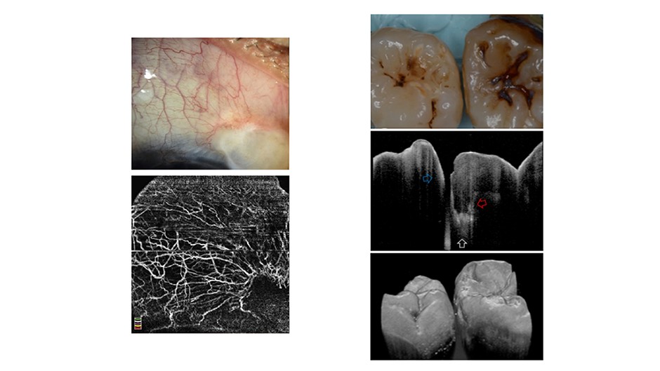
The imaging of tissues (inside or outside of the human body) could be a practical application of optical imaging. Tissue imaging is generally divided into two categories: soft tissues and hard tissues. As an example of soft tissue imaging, we can refer to corneal imaging with OCT (optical coherence tomography) method for drug delivery application. Imaging of soft tissues was much better and with higher resolutions than hard tissues due to deeper penetration and less scattering. However, researchers are recently able to record high-resolution images of bone and tooth tissues with current improvement in OCT or two-photon laser scanning.
 (Left) Optical imaging of corneal vascularization (soft tissue)-Ang et al. 2018. (Right) Optical imaging of teeth-Shimada et al. 2020
Light-activated therapy and surgery
Due to the energy contained in light, not only this source can be used to establish diagnosis methods, it can be used to develop treatment techniques for diseases such as cancer, poor eyesight, clogged arteries and so on. To get a clear picture of how optical methods treat the disease, it can be very useful to pay attention to the following applications:
I) Tissue engineering
Tissue engineering is a field of medical engineering that includes biocompatible artificial implants, tissue regeneration, tissue welding and soldering, and tissue restructuring and contouring. Light, especially lasers, is an efficient tool for the mentioned applications. The following are some of its applications in dermatology and ophthalmology:
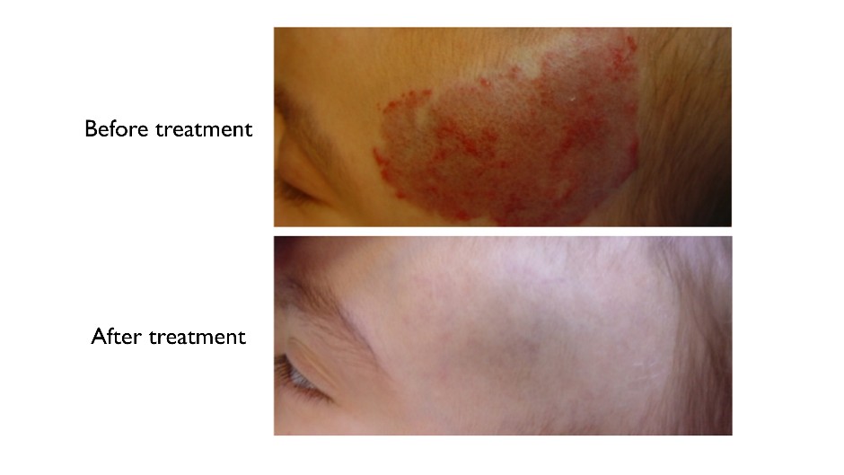
Dermatology: the treatment of vascular malformations, the removal of pigment lesions and tattoos, wrinkle removal, hair removal
 Laser treatment of vascular malformations-Jeremy et al. 2013
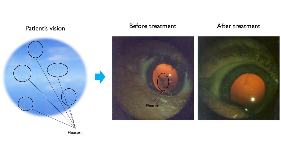
Ophthalmology: repair of blockage, leaky blood vessels, eye floater removal, reform the cornea for vision correction using the laser in situ keratomileusis (LASIK).
 Laser treatment of eye floaters - https://www.eyefloaters.com/eye-floater-types
II) Cancer therapy (photodynamic therapy)
It is a newly-found method in the treatment of cancer. It works by injecting a light-sensitive drug (called photosensitizers) into a patient. After three days, the healthy cells of the patient's body excrete the injected drug, while the cancer cells are still absorbing the drug. In the next stage, the body is exposed to light with a specific wavelength. This activates the drug in the cancer cell for the treatment purposes. One of the advantages of this method is that it does not require surgery.
Light has great capabilities in the development and advancement of medical science. The nature of light has led scientists, regardless of their expertise, to consider light as a suitable tool for improving the performance of conventional methods and even developing new methods to improve the quality of human life. Now, it might be a good time to think about what role light plays in our lives and how it can change our future...
About the author


