Illustrating Medicine
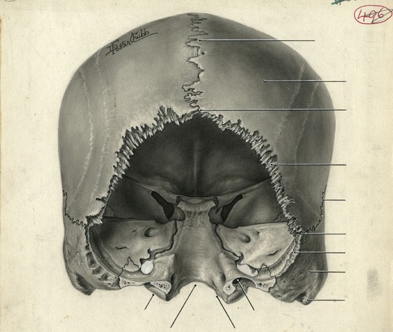
Illustrating Medicine features outstanding examples of the art of medical illustration in the 1940s, including carbon dust drawings by Dorothy Chubb, black and white water colours by Nancy Joy, and line drawings by Elizabeth Blackstock and Marguerite Drummond.
The exhibition highlights the skill of the illustrators, demonstrates the processes involved in image-making for an anatomical atlas, and exemplifies the key role of the medical illustrator in promoting regional anatomy as the vision of the body that became dominant in this period.
The exhibition is curated by Kim Sawchuk, a professor in Communication Studies, and Nancy Marrelli, Archivist Emerita at Concordia.
When: March 13, 2014 to May 1, 2014. Gallery hours: Monday to Thursday from 9 a.m. to 4:45 p.m. and Friday from 9 a.m. to 12:45 p.m.
Where: Media Gallery, Room 1.419, CJ Building, (7141 Sherbrooke St. W.), Loyola Campus.
The exhibition's organizers gratefully acknowledge their sponsors: the Social Sciences and Humanities Research Council of Canada; the Biomedical Communications Department-University of Toronto Mississauga; the Department of Anatomy, University of Toronto; the Department of Communication Studies, the Mobile Media Lab, Concordia University and the Media History Research Centre, Concordia University.

