Check out the winning photos from Concordia's 2023 Science Captured contest
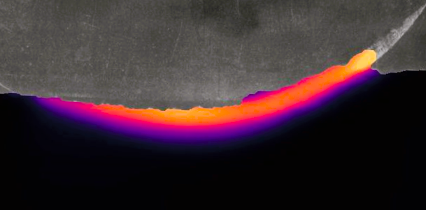
 “Soil Flow Beneath a Wheel in Lunar Soil Simulant”
by George Butt, MASc in Electrical and Computer Engineering
The School of Graduate Studies is celebrating this year’s winners of Concordia’s Science Captured Prize.
From brain cells to polar bears, the competition allows grad students to showcase their research through a vibrant image. Every year, four winners are selected to receive $250.
The photo above was taken by George Butt, MASc student in electrical and computer engineering. It shows a single-wheel testbed capturing subsurface soil movement in reduced gravity. "The colour gradient allows the soil flow speed to be visualized, where black is static and yellow is the highest speed."
Here are the other winning photos for 2023:

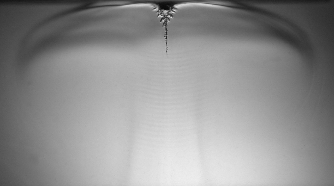
“Only the Sound Remains”
By Mohsen Habibi and Shervin Foroughi
Postdoc in mechanical engineering
Ultrasound waves create tiny oscillating air bubbles in a liquid polymer solution, transforming the liquid into a solid. "This new 3D printing method harnesses the power of sound waves and bubbles to create solid structures."

“Studying the ‘unseen microbial majority’ of the Arctic Ocean”
by Susan McLatchie
MSc, Biology
A polar bear treks across the expanse of the Arctic Ocean. "The Arctic Ocean's biodiversity, consisting mainly of microbes beneath the ice, is critical to the survival of the polar bear because it regulates the global climate."
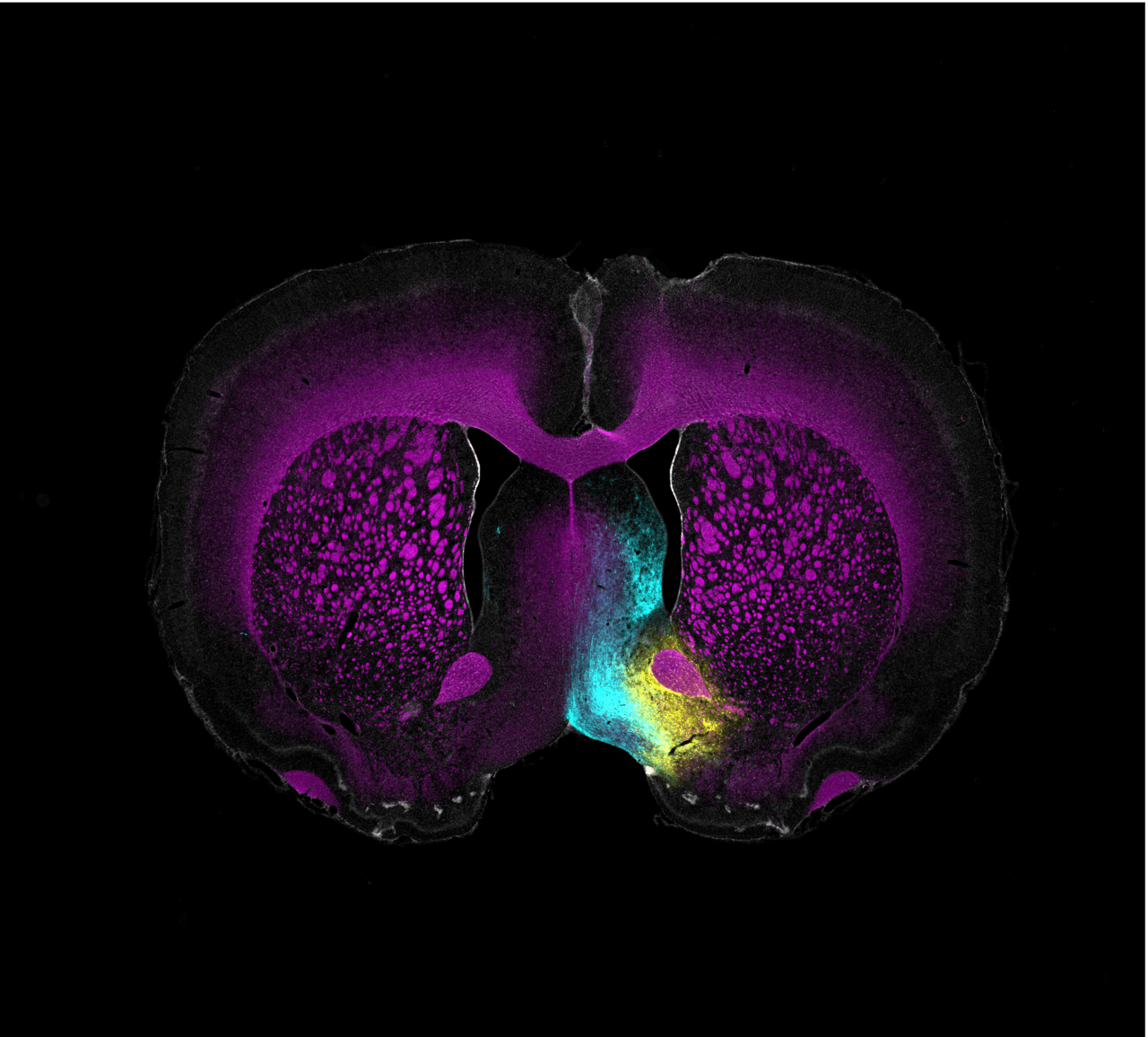
 “In Search of Reward Neurons in the Brain” by Jacques Voisard, MA Psychology
“In Search of Reward Neurons in the Brain”
by Jacques Voisard
MA, Psychology
This photo shows a tiny slice of a rat's brain. The parts coloured white are the cell nuclei. The thin, magenta layer around the nerve fibers is called the myelin sheath. The yellow lines show the path of special brain cells that start in the Diagonal Band of Broca. The cyan lines show the path of brain cells that start in the Lateral Hypothalamus.
Learn more about Concordia’s School of Graduate Studies.