Confocal microscopes
Nikon SR-SD

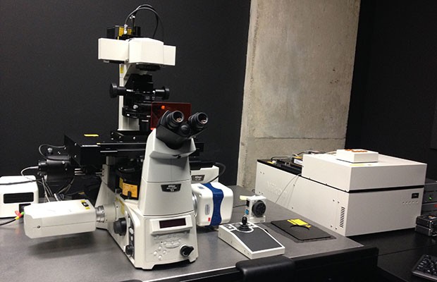
The Nikon Super-Resolution Spinning Disk (SR-SD) is based on a Nikon Ti2 microscope equipped with a motorized XY stage, Piezo-actuated Z-stage, 4x, 10x, 20x, 40x, 60x and 100x lenses, a Yokogawa CSUX1 Spinning Disk head, GATACA Live-SR unit, Andor Zyla 4.2 sCMOS camera (2048x2048 pixels), and an LCI stage-top incubator for live-cell imaging. Fluorescence excitation is provided by 405/488/561 and 638nm lasers, with emission filters for DAPI+Far-Red/GFP/Texas Red/Far-Red and a 650nm longpass. The system is operated using µManager software.
Example images from SR-SD
Quorum/CREST Optics Cicero

The Cicero spinning disk is a high-speed lenseless spinning disk system allowing rapid confocal imaging of living samples. The system is based on a Zeiss Axio Observer microscope equipped with motorized XYZ stage, 5x, 20x, 40x, 60x and 100x Plan-Apo lenses, a Hamamatsu Flash 4.0 LT camera (2048x2048 pixels) and an environmental chamber for long-term live imaging. Fluorescence excitation is provided by the 89North LDI-5 laser launch, with lines at 405/488/555/640nm, with emission filters for DAPI, Alexafluor488, Alexafluor 568 and a longpass far-red filter. The system is operated using Quorum's Volocity software or µManager.
Images from the Cicero Spinning Disk
Nikon C2 laser scanning confocal microscope

The Nikon Laser scanning C2 system is a fully motorized inverted microscope, equipped with four lasers (405, 488, 561, 640 nm), for confocal imaging, bright 20x, 40x, 60x and 100x objective lenses; DIC optics, a motorized stage and a Nikon Perfect Focus autofocus system. The system is operated using NIS-Elements software.
Images from the Nikon C2 Confocal
Olympus Fluoview FV10i Laser scanning microscope

The Fluoview FV10i (or "confocal-in-a-box") is a self-contained confocal laser scanning microscope. It is outfitted with four laser diodes (405nm, 473nm, 635nm, 559nm) and uses a photomultiplier-based spectral system for detection. It has a 10× air and a 60× oil immersion objectives lens with the possibility to zoom up to 10 times to provide highly resolved phase contrast and fluorescent confocal images (up to 0.027 μm/pixel).
The FV10i is equipped with an automatic detection of interface between specimen and cover glass by laser reflection light detection. This scope is suitable for imaging of fixed cells or tissues.


