Feature: Your Next Doctor is an Engineer

January 20, 2015
|
By Laurence Miall


We are now on the verge of what has been described by Nobel Laureate Phillip Sharp as a “third revolution” in the life sciences, in which interdisciplinary collaboration among engineers and physical and computational scientists promises to unleash yet more innovations. Concordians are playing their part.
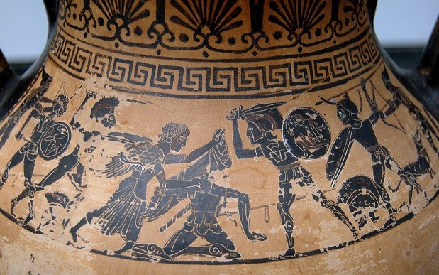
Aphrodite rescuing her son Aeneas wounded in fight, scene from The Iliad. Photo by Bibi Saint-Pol (Own work) [Public domain], via Wikimedia Commons

According to a modern-day analysis of the 10-year Trojan War, which was waged sometime between the 11th century and 12th century in ancient Greece (and immortalized by Homer’s famous poem The Iliad), the death rate from arrow wounds was 42 percent, slingshot wounds, 67 percent, spear wounds, 80 percent, and sword wounds a staggering 100 percent. Many injuries were nothing less than a death sentence. Centuries later in the Vietnam War, the mortality rate for American soldiers who had suffered abdominal wounds was only 4.6 percent. By this point in history, any conventional army was typically followed by a second, smaller army as well versed in saving lives as their counterparts were in destroying them. Doctors, nurses and surgeons benefited from the latest medical equipment, and helicopters could rapidly convey the wounded from the battlefield to the bed.

From antiquity to the 20th century, a huge array of technology advances had radically altered, not just war medicine, but all of the health sciences. Engineers were behind many of the improvements. We are now on the verge of what has been described by Nobel Laureate Phillip Sharp as a “third revolution” in the life sciences, in which interdisciplinary collaboration among engineers and physical and computational scientists promises to unleash yet more innovations. Concordians are playing their part.

Bone Scaffold

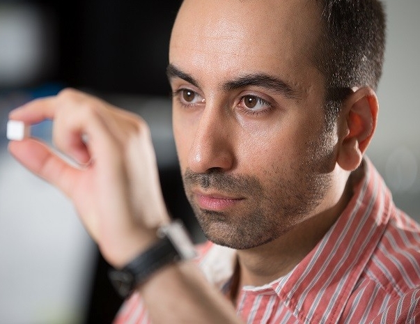
The fate of Americans in Vietnam is a reference point for Ehsan Rezabeigi, an Iranian-born international student who came to Concordia to work on his doctorate. The war saw the widespread use of bone substitutes, he explains. These were typically metal bars, inserted into the body to help heal broken limbs. This practice is still the norm today, but it has numerous disadvantages. The surgery required is highly invasive, and the metal prostheses are heavy and cumbersome.

Under the supervision of professors Paula Wood-Adams, who is also Concordia’s Dean of Graduate Studies, and Robin Drew in the Department of Mechanical and Industrial Engineering, Rezabeigi is hard
at work on a technology that could revolutionize the treatment of bone fractures.

He is developing in three distinct phases a lightweight implant that can be surgically inserted into the body to support and promote bone regrowth.
Phase one involved synthesizing bioactive glass particles that are capable of feeding bone cells and helping them to grow. In phase two, he developed a novel technique of creating a highly porous polymer foam (a polymer is a structure made up of many molecules in a long chain). This polymer foam has high-performing mechanical properties: it’s strong, and it can be used in load-bearing parts of the body like the shoulders and knees. The third phase of Rezabeigi’s research consists of combining the bioactive glass particles with the polymer to make a brand-new composite material: a bone scaffold.

“The ideal scaffold has the same properties as bone. This is the goal we’re striving toward,” Rezabeigi says. The scaffold must be highly porous to allow for vascularization — the formation of blood vessels. This is, in part, what allows for bone growth. Meanwhile, small pores in the scaffold allow for cell attachment.

One of the most astonishingly futuristic properties of the bone scaffold is that it’s bio-reabsorbable. In other words, it can safely dissolve into the body over time once it has completed its mission.

[Watch a short video about Rezabeigi’s research]

Rezabeigi is grateful to the Natural Sciences and Engineering Research Council (NSERC) and Concordia for their support of this project.

Simulated Surgery and Touch

Operating on a patient is one of the most difficult and painstaking skills required for a medical practitioner. From the patient’s standpoint, it can be an ordeal: recovery from surgery can take weeks, even months, and opening up the body introduces the risk of infection. It’s no surprise, then, that what is called minimally-invasive surgery has become popular. This involves making keyhole incisions in the body and performing the operation using laparoscopic tools, which are long rod-shaped devices of only five millimetres in diameter or less, equipped with some form of visualizing device that allows the surgeon to remotely view what she is operating on.

Increasingly, minimally invasive surgeries are carried out with the help of robots, whose artificial appendages can be even more precise and dependable than a surgeon’s hands.

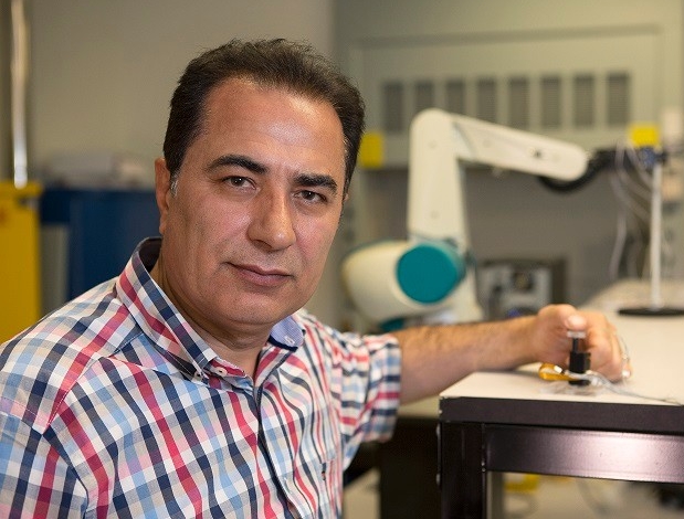
Minimally invasive surgery is Javad Dargahi’s specialty. Not the surgery itself, but the refinement of devices and techniques to help. The mechanical engineering professor has devoted a lot of his research to haptic feedback — bringing the human touch back to robot surgery.

“What is missing with robot surgery is tactile feedback,” Dargahi explains. “Haptic feedback involves collecting tactile information such as texture and hardness, and kinesthetic information about certain forces from the limbs, and then playing this back to the surgeon.”

Among numerous projects, Dargahi has helped to devise and test sensors integrated into a catheter that can be inserted into a patient’s body. The sensors relay information about the hardness and softness of tissues. The information helps a surgeon to determine where to place the anchor required for a complete mitral valve replacement during heart surgery. The sensors have so far proven successful in a simulated environment

Dargahi has also made great strides forwards in the related field of surgery simulation. What kinds of movements does a skillful surgeon make? Surgeons in training can benefit from first-hand knowledge of this, and Dargahi and his students are developing systems composed of cameras and sensors to measure surgeon’s movements and compare them against those of trainees in a simulated surgery environment.

Dargahi is grateful to the Natural Sciences and Engineering Research Council (NSERC) for a Discovery grant in support of this research.

Lab-on-a-chip

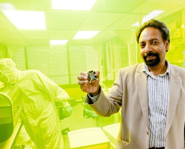
Muthukumaran Packirisamy is also a mechanical engineering professor, but working in a radically different field. You’d need a powerful microscope to properly understand his diverse contributions to the life sciences. He has found multiple applications for what is call lab-on-a-chip technology. Nanotechnology has permitted complex experiments to be enacted on chips no bigger than your fingernail. It’s a technology with diverse applications.

For example, Packirisamy developed a chip capable of detecting growth hormone in milk. Growth hormone is banned in Canada. Before lab-on-a-chip technology, a farmer’s only option for identifying the hormone was to send samples to laboratories hundreds of kilometres away and wait days to get back results. Thanks to Packirisamy’s patented kit, tests can be conducted right on the farm, getting results in minutes.

“The kit can trace hormone in quantities as minute as two nanograms per millilitre. Previously we could only measure 100 nanograms per millitre,” Packirisamy explains.(A nanogram is one billionth of a gram.)

Packirisamy is currently working with Nahum Sonenberg, a microbiologist and biochemist at McGill University’s Goodman Cancer Research Centre. This time Packirisamy’s lab-on-a-chip technology is being applied to conducting tests on liver cancer cells. This kind of experimentation holds a lot of promise, since implanting cells in a chip can often be an alternative to implanting them in rats or other animals, and can also significantly speed up the
testing time.

“The microfluidic environment inside the chip is for our purposes better than a Petri dish,” he says. “You can simulate all sorts of environments. In a healthy environment, the cells grow; in a toxic environment,
the cells die. We custom design the chip for each application — this definitely isn’t a one-size-fits-all invention.”

Packirisamy is grateful for the support of the following: Natural Sciences and Engineering Research Council (NSERC), Concordia University Research Chairs program, Fonds de recherche Nature et technologies (FRQNT), Ministère de l'Économie, de l'Innovation et des Exportations (MDEIE), Canadian Foundation for Innovation, and industry partners.

"Seeing" the Invisible

When you open up somebody’s skull in order to operate, the brain can shift by as much as four centimetres. Known as brain deformation, it’s one of the trickiest problems for neurosurgeons. A newcomer to Concordia, computer scientist Hassan Rivaz, has dedicated himself to solving this and several other complex problems in the field of medical image processing.

Rivaz says he was first drawn to medical applications of engineering because, as he puts it, “I wanted my research to have humane applications.” He made a switch from mechanical engineering to computer science, and subsequently earned a PhD at Johns Hopkins University, largely out of an interest in elastography. This is a technique of mapping parts of the body using medical imaging, focusing on the elasticity of tissue, which provides clues as to possible disease. Finding very hard tissue, for example, can indicate the presence of a cancerous tumour that can be invisible in ultrasound.

To return to the open skull. When a neurosurgeon is operating, she will be referring to an MRI of the brain conducted earlier. But if the brain has now deformed, this map is no longer accurate. This is where Rivaz’s algorithms can help point the way. During his postdoctoral fellowship at the Montreal Neurological Institute, Rivaz developed algorithms that would do image registration—this involves super-sophisticated math that can align the pre-operation image with the images produced during the surgery itself.

“This doesn’t require any new hardware beyond ultrasound,” explains Rivaz (ultrasound being the main mapping tool used in the operating room.) “You just need software, which is both convenient and inexpensive.”

This is an example of an intervention — the use of Rivaz’s research during an actual medical procedure. His research has also had applications in diagnostics, proving to be extremely powerful.

Rivaz is grateful for the support of the following: Natural Sciences and Engineering Research Council of Canada (NSERC), Jeanne Timmins Costello Fellowship, Department of Defense (DoD) and the Link Foundation.

Convergence

“An individual who becomes educated in the current technical and biomedical world will have just a multitude of opportunities to use their education and their talents,” Phillip Sharp said in an interview just a few short years ago. The fruitful collaborations at Concordia and beyond prove the power of convergence, the term Sharp uses to describe the happy marriage of engineering, physical and life sciences.

Rivaz’s latest collaboration is with Paul Martineau, a sport medicine researcher at Concordia’s PERFORM Centre. They’ll be figuring out potential improvements for diagnosing muscle and tendon injuries. Packirisamy, meanwhile, has shipped one of his sensors to India where it has flown aboard a plane in order to detect harmful ammonia in urban areas. Dargahi continues his collaboration with colleagues at Montreal’s Jewish General Hospital and is celebrating the publication of his third book. And Ehsan Rezabeigi, progressing with his PhD, is explaining his research to scholars around the world through recently co-authored publications in Materials Science and Engineering and Polymer, and has plenty more publications in store.

If Sharp’s Third Revolution is indeed nigh, these four men are among the first at the barricades.


Back to top

© Concordia University