Concordia prof helps augment reality for surgery

Marta Kersten-Oertel is developing visual navigation tools to assist doctors in the operating room
March 19, 2018
|
By Joseph Leger


Spatial visualization is an essential skill surgeons have to master to perform even the most basic surgical procedures. Surgeons routinely have to navigate complex, three-dimensional spaces without damaging vital tissue, nerves or organs to reach an area requiring medical attention.

Marta Kersten-Oertel, assistant professor in Concordia’s Department of Computer Science and Software Engineering and head of the Applied Perception Lab, is developing navigational tools to help increase precision and accuracy in the operating room.

WATCH: Marta Kersten-Oertel specializes in developing and testing visualization, display and interaction methods in the context of image-guided surgery. In this video she describes her research and its applications at the Concordia Chancellor’s Builders Circle and Friends Dinner. 

“I am working on software tools to visualize and display anatomical patient images to help guide surgeons to treat neurological disease,” Kersten-Oertel explains. “I look at how to visualize and add depth to medical images. One of the applications of my work is in image-guided neurosurgery or neuronavigation.”

Currently, surgical visualization aids are displayed on monitors, forcing surgeons to cycle their focus between the surgery and the visual guide. To bypass this, Kersten-Oertel projects images directly onto the surgical area — effectively superimposing an accurate, three-dimensional anatomical map to act as a guide during invasive medical procedures.

“Think about how most of us get around by car,” she says. “We navigate with the help of GPS. The danger is that by having to look at a separate navigation system, you shift your focus away from the road. A few years back, BMW developed a solution by introducing augmented reality visualization. The navigation information is displayed directly on the car windshield, similar to a fighter pilot’s heads-up display.”
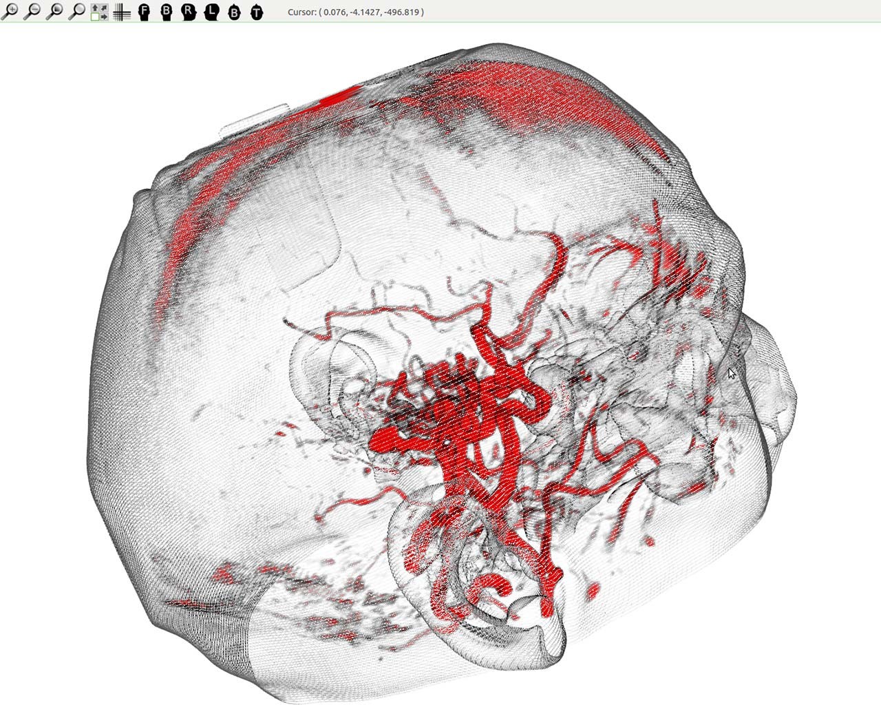
Three-dimensional images projected onto a skull Three-dimensional images projected onto a skull will provide a sense of depth, accurately mapping the internal anatomy. | Photo: Marta Kersten-Oertel
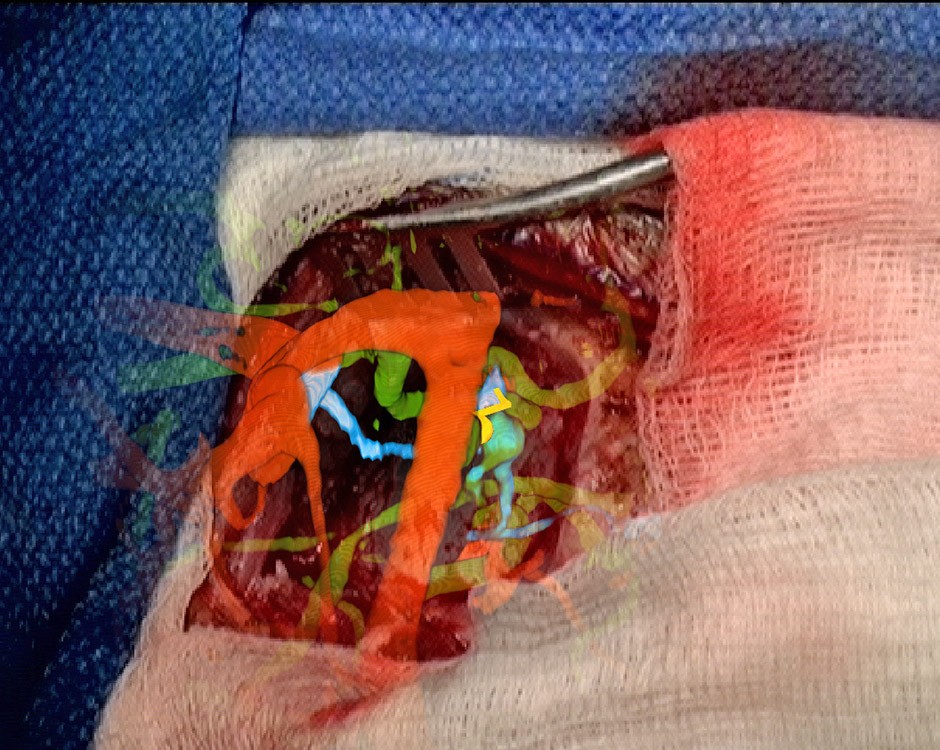
Detail showing surgical area Images are superimposed onto the surgical area, providing detailed guides for surgeons. | Photo: Marta Kersten-Oertel

Kersten-Oertel’s work uses the same type of navigation technology in the operating room, where infrared cameras track surgical tools to help surgeons navigate. Similar to BMW’s navigation system, she is merging three-dimensional models of the patient anatomy and surgical plans, enabling surgeons to locate and target disease with greater precision and preserve more healthy tissue.

To develop her methods and validate her results, Kersten-Oertel works closely with researchers and clinicians at the Montreal Neurological Institute and Hospital.

Inspired by art

Kersten-Oertel says her work is inspired from her time living in Vienna, Austria, where she indulged her passion for fine art by exploring museums. She was particularly interested in how art evolved through different periods.

“In the Gothic period, paintings were flat and lacked depth,” Kersten-Oertel says. “By the early Renaissance, artists began applying perspective rules to make an image on a flat canvas appear three-dimensional. This idea of displaying images so that they look three-dimensional has very much impacted my research.”

She was so fascinated by this evolution of art that she earned a minor in art history while pursuing her BSc at Queen’s University in Kingston, Ont. Luckily for the medical world, Kersten-Oertel decided to pursue a career in computer science and software engineering instead.

“I want to move this research forward for the benefit of society, so that these technologies will one day become a mainstay of the operating room,” she says.

#CUpride

Related links


Back to top

© Concordia University