Researchers are improving how brain-imaging data is collected and visualized
Hacking into people’s minds like they do in The Matrix movies may seem far-fetched, but improved neuroimaging techniques are making it easier to unlock the mysteries of the human brain.
There are many different ways to gather images of the brain. Some — such as magnetic resonance imaging (MRI) — create structural images that show the organ’s anatomy. MRIs are used primarily to diagnose large-scale problems, such as tumors.
Another group of imaging methods produce functional images, which allow scientists to observe processes of activation in the brain. For example, functional images can detect which part of the organ is activated during certain motor tasks.
Near-Infrared Spectroscopy (NIRS) falls into this latter category. It's used to investigate changes in the concentration of oxygen saturation in hemoglobin on the cortex. Two researchers at Concordia’s PERFORM Centre want to improve the quality of data gathered by this method.
Zhengchen Cai, a PhD candidate in Physics, and Thomas Vincent, a postdoctoral fellow in the Department of Physics, have been have been working on methodology and software development for NIRS data analysis over the last few years.
Together with Cai’s supervisor Christophe Grova, an associate professor in Concordia’s Department of Physics, they will lead a workshop on May 14 and 15 during PERFORM Centre Research Week showcasing the new software they have created.
'Neuroimaging is a highly multidisciplinary field'
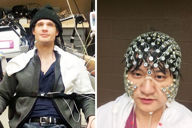
How do these images (above) relate to your research at Concordia?
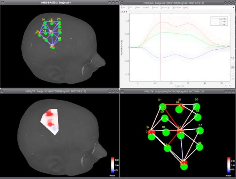
Thomas Vincent: The above, left image shows me with the NIRS device attached to my head. The sensors are hidden under a cap because they're sensitive to ambient light. The near-infrared light penetrates through the skull.
Hence, this neuroimaging device is able to track variations of blood oxygenation at the level of the cortex. These variations reflect cerebral activity, for instance here during a motor finger-tapping task.
The second image (top) illustrates my software development work — namely the NIRSTORM toolbox — dedicated to processing NIRS data. It shows the activity in the brain’s motor area that is being caused by finger tapping.
Zhengchen Cai: To investigate the hemodynamic activities of the human brain using NIRS, we must collect data according to a specific experimental protocol. The equipment shown in the image below is the Brainsight NIRS system, which “illuminates” the brain with near-infrared light.
It also detects light intensity variations induced by changes in hemoglobin concentration in the brain. The Brainsight system — along with anatomical MRIs of the subject — act like a map of the brain.
By using them, we can precisely probe sensors on top of the area we’re interested in or stimulate a specific region of the cortex.
What is the hoped-for result of your project? And what impact could you see it having on people's lives?
TV: The main advantage of NIRS is its ability to work wirelessly, using a small device that can be carried on a belt for example.
This enables freedom of movement and testing of human behaviour in a natural context. For instance, an interesting application I'm involved in is studying a memory task while walking. This allows for the study of divided attention and its underlying cerebral mechanism. It also has a potential application in the prevention of falls in older populations.
ZC: With the NIRS reconstruction algorithm developed in our lab, we expect to be able to recover the locations, sizes and amplitudes of the hemodynamic responses underlying the scalp and their spatial extent along the cortical surface.
By applying this technique with transcranial magnetic stimulation — which temporarily modifies the level of excitability in the brain — we would like to characterize the relationship between the task-related hemodynamic responses and cortical excitability.
Most importantly, this project is a necessary first step to achieve physio-pathological insight into the relationship between excitability and hemodynamic activity. This will help us treat patients with epilepsy in the future by offering applications of non-invasive brain stimulation.
 The Concordia researchers evaluated their software using data acquired from a Brainsight NIRS device (pictured). | Image courtesy of Rogue Research Inc.
What are some of the major challenges you face in your research?
TV: The main challenge I'm concerned with is data quality and experimental noise. To address this, our efforts focus on improving the measurement technique by finding the optimal way of placing the sensors. Moreover, I develop advanced statistical methods to better distinguish cognitive-related components from the noise sources in the signal.
ZC: My research project consists of performing comprehensive tasks and taking neuroimaging studies through every step of the scientific process — from experiment design and subject recruitment to methodology development and data analysis. I need to be able to handle both the practical and theoretical work this requires.
What person, experience or moment in time first inspired you to study this subject and get involved in the field?
TV: When I joined Christophe Grova’s and Louis Bherer’s research teams, I discovered a richer way of doing neuroimaging which was grounded in real-life experiments. I could develop advanced statistical models while also participating in concrete experimental setups. On the application side, I could take part in the prevention of cognitive decline in older populations.
ZC: Everything fell into place naturally. I did my bachelor’s degree in electronic engineering and it related to the research work that I'm doing now. If there's one far-fetched inspiration, I guess it's because I'm a big fan of The Matrix — it would be fantastic to investigate the human brain like that. Since we are modifying the excitability level of the cortex, it's like a mild “hacking” of the brain.
How can interested STEM students get involved in this line of research? What advice would you give them?
TV: Neuroimaging is a highly multidisciplinary field involving physics, cognition, electrical engineering, advanced mathematics and statistics, as well as software development. The best advice to give to students is have expertise in at least two of these topics.
What do you like best about being at Concordia?
TV: I'm especially impressed by how Concordia creates new innovative research paradigms. The PERFORM Centre is one striking example of a unique platform that brings together high-end neuroimaging techniques, a biochemical laboratory and a nutrition platform, as well as physical exercise research equipment.
ZC: It is a great opportunity to study in Christophe Grova’s lab at Concordia because we often connect with experts in the field in order to learn and share our experiences. Thanks to the PERFORM Centre, we have a nice platform to study neuroimaging. It also helps us build a research community in this field at the university.
Are there any partners, agencies or other funding/support attached to your research?
TV: Through the grants of Christophe Grova and Louis Bherer, my work received funding from the Natural Sciences and Engineering Research Council of Canada (NSERC), the Canadian Institutes of Health Research (CIHR) and the Concordia University Research Chair in Preventive Health Research.
ZC: My project is supported by a Quebec Bio-imaging Network grant provided by the Fonds de recherche du Québec – Santé and an NSERC Discovery Grant.
Find out more about the NIRSTORM workshop and the PERFORM Centre Research Week.