Christophe Grova, Ph.D.
PC-2.147
(514) 848-2424 ext. 4221
Email: christophe.grova@concordia.ca
Grova Research Group
The Multimodal Functional Imaging Laboratory, directed by Dr. Christophe Grova, is a multidisciplinary team composed of neurologists and methodologists. The laboratory is actually based on two sites:
- Biomedical Engineering Department and part of the epilepsy group of the Montreal Neurological Institute, McGill University
- Department of Physics and School of Health, Concordia University
We investigate multimodal data fusion to characterize brain mechanisms and especially epileptic activity. Our research project aims at developing methods to appropriately combine multimodal data in order to detect additional information that could be missed by considering each modality individually. A typical challenge is to combine modalities directly measuring neuronal activity with high temporal resolution with other modalities indirectly measuring the same function with high spatial resolution, through hemodynamic processes for instance. The project will involve the integration of three promising functional modalities:
- Simultaneous ElectroEncephaloGraphy (EEG) - MagnetoEncephaloGraphy (MEG) acquisitions, measuring directly on the scalp electric and magnetic components of signals generated by neurons synchronously active (at a ms scale).
- Simultaneous EEG - functional Magnetic Resonance Imaging (fMRI) acquisitions to measure, within the whole brain at a second scale, hemodynamic responses that correlate with signals detected on scalp EEG.
- Simultaneous EEG - Near InfraRed Spectroscopy (NIRS) acquisitions to measure local changes in oxy- and deoxy-hemoglobin at the time of signals detected on scalp EEG, by exploiting absorption properties of infrared light within brain tissues using optic fibres placed on the surface of the head. Note that our EEG/NIRS laboratory is located in Biomedical Engineering dpt and easily accessible from the McConnell Brain Imaging Centre.
The principal clinical application of this project will be to combine these three modalities using multimodal data fusion techniques to characterize brain regions involved during epileptic activity.
Multi FunkIm Research
Using cutting-edge non invasive methods involving simultaneous EEG/MEG, EEG/fMRI and EEG/NIRS recordings, our objectives consist in proposing:
Theme 1: EEG/MEG source localization
We propose a methodology to localize along the cortical surface, electrophysiology data obtained at a millisecond time scale from scalp recordings. This results in solving the so-called inverse problem of source localization. Our main goal consists in the developpment of Maximum Entropy on the Mean (MEM) source localization, in the context of EEG source imaging, MEG source imaging and also EEG/MEG fusion, as the only distributed source localization method able to recover the spatial extent of the underlying generators.
This video shows the steps and the results of Electroencephalography (EEG) source localization for a visual half field paradigm. EEG is a technique that measures the neuronal activity in the brain using electrodes that are pasted on the scalp. It can measure brain signals at every millisecond, however, without advanced mathematical and physical tools it could not provide 3-dimensional (3D) information on the brain. Our research involves fusing the EEG signals -measured with a high-density EEG system (256 electrodes, EGI)- with the 3D anatomical information of the brain, obtained with Magnetic Resonance Imaging, to localize the neuronal activity on the cortex.
Complete multimodal investigation involving MEG source localization, fMRI response and intra-cranial EEG investigation.
Theme 2: Volumetric NIRS
Spatio-temporal reconstruction of oxy-hemoglobin (HbO) and deoxy-hemoglobin (HbR) responses. We propose a strategy from EEG/NIRS data acquisition, signal detection to volumetric reconstruction to demonstrate the ability of NIRS to localize, in space and in time, fluctuations of HbR and HbO.
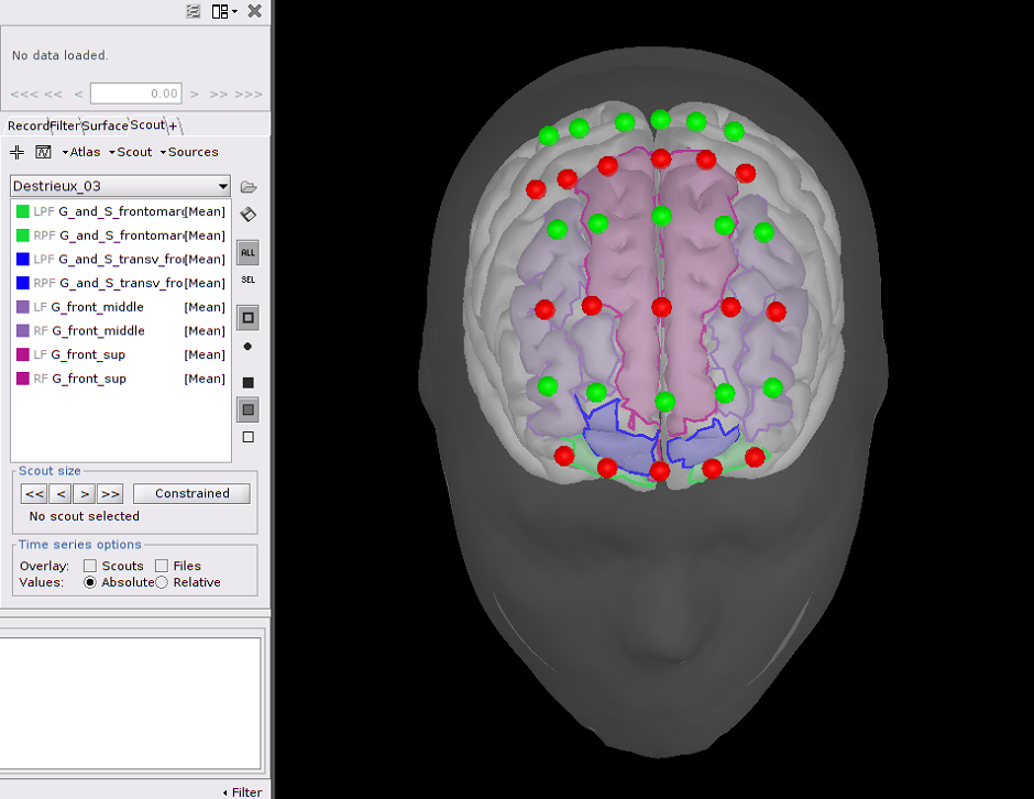 Frontal NIRS montage used for a working memory experiment where optodes are depicted as spheres, with green ones being detectors and red ones emitters. A transparent template head mesh is shown in dark grey, as well as a template cortex in light grey. Finally, an atlas of the pre-frontal cortex is depicted in purple (superior prefrontal), blue (transversal frontal) and green (orbito-frontal).
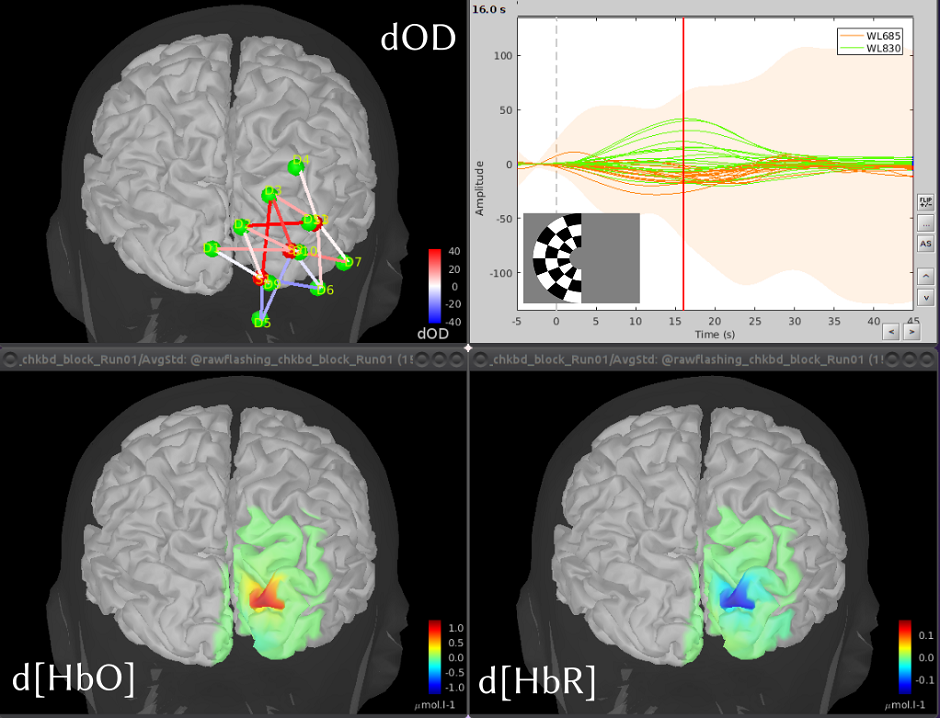 Coritcal reconstruction of the variations of oxy (HbO) and deoxy (HbR) hemoglobin concentrations, evoked by the visualization of flashing checkerboards. The map on the top left depicts the optimal montage placed on the primary visual cortex. The bottom pictures show reconstructed maps of the maximum evoked HbO and HbR variations. The graph with the time courses shows the temporal hemodynamics variations, evoked by the visual stimulations.
Theme 3: Multimodal characterization of Neurovascular Coupling (NVC) during normal and pathological conditions
Multimodal characterization of Neurovascular Coupling (NVC) during normal and pathological conditions using (i) EEG/MEG sources to model neuronal bioelectrical input and (ii) fMRI and NIRS data to monitor brain hemodynamic response. NVC is the main principle allowing indirect measurement of brain activity (in fMRI, NIRS or Positron Emission Tomography) exploiting processes linking neuronal activity and brain vascular response. However, these processes are not well-known, especially in pathological conditions. Our objective is to combine multimodal non-invasive data to shed light on NVC processes.
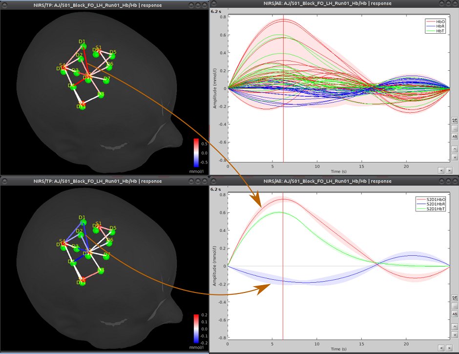 Analysis of functional Near-InfraRed Spectroscopy (fNIRS) data acquired during a motor experiment. fNIRS is a technique that measures brain activity through the absorption of near infrared light by the cerebral hemoglobin (Hb). Hb concentration indeed varies locally when there is local neuronal activity. This is the so-called neuro-vascular coupling. The curves on the right show the variations of concentration of oxy- (red) and deoxy- (blue) hemoglobin evoked by a finger tapping task. On the left part, the amplitudes of these variations are displayed, mapped onto each measurement pair of optodes.
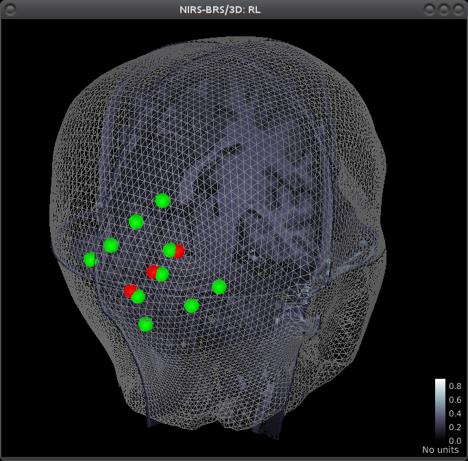 Multimodal view of a NIRS occipital montage. The head mesh segmented from the anatomical MRI is shown as a grey wireframe. Slices of the subject’s anatomy are shown in greyscale. The NIRS optode placement is depicted as spheres, with green ones being detectors and red ones emitters.
Theme 4: Multimodal functional connectivity (FC) characterizing resting state data in normal and pathological conditions
While resting state fMRI data have exhibited regions showing distant correlations at low frequency (<0.05Hz) organized as connected networks, the consistent Resting State Networks (RSN), our objective is to combine multimodal data to identify EEG/MEG signatures that would explain such network organization and to characterize pathological networks at the individual level, as potential disease biomarkers.
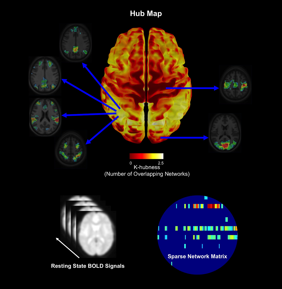 The organization of brain networks can be studied using the blood oxygen-level dependent (BOLD) signals measured using functional magnetic resonance imaging (fMRI) when a subject is not performing an explicit task (resting-state). We proposed a method called SParsity-based Analysis of Reliable K-hubness (SPARK) to model and estimate “hubs” of functional networks by measuring K-hubness in each voxel with good reliability of the estimation at the individual level. Hubs are defined as voxels that are involved in multiple (a sparse number K>1) networks, promoting inter-network connectivity and global communication that are related to higher-level brain functions.
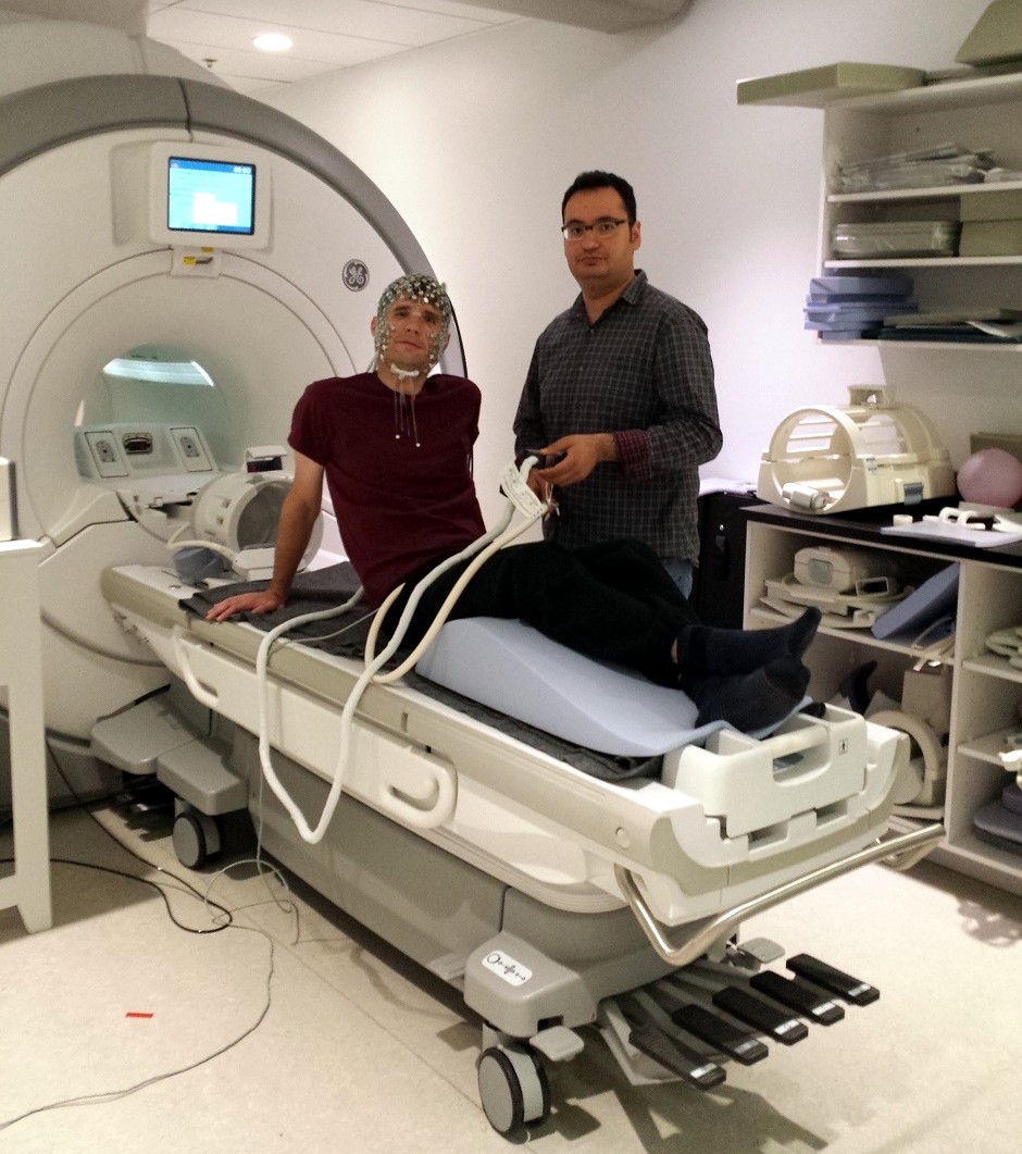 Joint acquisition of electroencephalography (EEG) and functional Magnetic Resonance Imaging (MRI). A high density EEG cap is placed on the participant’s head before he enters the MRI scanner in the back.
Multi FunkIm Members
Principal investigator

Collaborators

Pierre Bellec, Ph.D.
Simexp lab: laboratory for brain simulation and exploration
Phone: (514) 340 3540 ext.3367
Fax: (514) 340 3530
Email: pierre.bellec@criugm.qc.ca

Habib Benali, Ph.D.
Scientific Direction of the PERFORM Centre, Concordia University
Phone: (514) 848-2424 ext. 7597
Email: habib.benali@concordia.ca
Research Interests: Human brain computational modeling and functional connectivity analysis. Multimodal analysis of electromagnetic and hemodynamic processes in the brain and spinal cord.

Thanh Dang-Vu, M.D., Ph.D.
Sleep, Cognition and Neuroimaging Laboratory (SCNLab)
Phone: (514) 340-3540 ext. 3991
Email: tt.dangvu@concordia.ca
Research Interests: Sleep pathologies. Biomarkers in neuroimaging in sleep associated to aging. Neurodegenerative diseases and cognitive decline.

Birgit Frauscher, M.D., Ph.D.
ANPHY Lab, McGill University
Phone: (514) 398 1952
Email: birgit.frauscher@mcgill.ca
Research Interests: Development of novel seizure-independent EEG markers for the epileptogenic zone in for accurate diagnosis of epilepsy. Investigation of the relationship between sleep and epilepsy. Brain physiology during wakefulness and sleep.

Claudine Gauthier, Ph.D.
Quantitative Physiological Imaging Lab, Department of Physics, Concordia University
Phone: (514) 848-2424 ext. 2193
Email: claudine.gauthier@concordia.ca
Research Interests: Cerebral metabolic and vascular imaging. The impact of aging and lifestyle on the brain. Functional MRI. BOLD signal modeling. Quantitative imaging of plasticity.

Jean Gotman, Ph.D.
Gotman Lab
Phone: (514) 398 1953
Fax: (514) 398 8106
Email: jean.gotman@mcgill.ca
Research interests: Multi-modal analysis of interictal events using EEG, fMRI, source-localization and dipole modeling, SEEG, SPECT.

Eliane Kobayashi, M.D., Ph.D.
Epilepsy Research Group, Montreal Neurological Hospital
Phone: (514) 398 6644, ext 00469
Fax: (514) 398 8106
Email: eliane.kobayashi@mcgill.ca

Jean-Marc Lina, Ph.D.
Centre d’études avancées en médecine du sommeil (CÉAMS), Université de Montréal
Phone: (514) 396 8688
Email: jean-marc.lina@etsmtl.com
Research interests: Wavelets, statistical modeling and brain imaging, algorithms for statistical learning.
Research associates
Tanguy Hedrick
Email: tanguy.hedrich@gmail.com
Education
· Ph.D. in Biomedical Engineering, McGill University, Montréal, 2019
· Master's in Biomedical Engineering, McGill University, Montréal, 2013
· Bachelor’s in Computer Engineering, University of Technology of Compiègne, France, 2011
Current research
His work is focused on source imaging applied to EEG and MEG, in the context of epilepsy. In 2019, he received a Ph.D. in Biomedical Engineering with a thesis entitled: "Combining high-density electrical source imaging and hemodynamic responses to epileptic discharges". He then obtained a position as a research associate with the collaboration of Dr. Frauscher and Dr. Grova.
Postdoctoral fellows

Chifaou Abdallah, M.D.
Email: chifaouah@gmail.com
Education
Chifaou Abdallah is a neurologist specialized in epilepsy and neurophysiology. She did her medical training at Pierre & Marie Curie University in Paris, her residency in neurology and clinical fellowship in epilepsy at the University Hospital of Nancy and the Lorraine University in France. Then, she joined Dr Christophe Grova’s lab at McGill University.
Current research
· McGill University (current)
Her research activity is focused on two projects related to epilepsy:
● Clinical value of magnetoencephalography and EEG/fMRI in the presurgical workup of patients with drug-resistant epilepsy, comparison with intracranial EEG finding
● Investigation of the relationship between sleep patterns and neurovascular coupling processes occurring at the time of epileptic discharges, using simultaneous EEG-Near Infrared Spectroscopy recordings.
Research interests
Using a multimodal approach (magnetoencephalography, EEG-HD, fMRI), her research interest is mainly to elaborate criteria for a better understanding and use of these non-invasive tools in order to improve the postsurgical outcomes and to select good patients for epilepsy surgery.
Hassan Khajehpour
Email: h-khajehpoor@alumnus.tums.ac.ir
Education
· Ph.D. of Biomedical Engineering, Department of Medical Physics and Biomedical Engineering, School of Medicine, Tehran University of Medical Sciences (TUMS)
Current research
· Department of Physics, Concordia University (current).
There is a surgical solution for patients with drug-resistant epilepsy, but the surgical outcomes are not always promising because many patients are not still seizure-free. He is working on a project, supervised by Prof. Christophe Grova, aimed to predict surgical outcomes in seizure patients by multimodal imaging: MEG/EEG, MRI, and iEEG. The approach is based on wavelet-based Maximum Entropy on the Mean to source localization followed by connectivity analysis predicting surgical outcomes.
Research interests
He is interested in research characterizing brain disorders using multimodal imaging aimed to discover new data-driven treatments. He is also interested in projects aimed to investigate brain stimulation impact, like tDCS, on drug craving.

Makoto Uji
Email: makoto.uji@concordia.ca
Education
· Ph.D., School of Sport and Exercise Sciences, Liverpool John Moores University
Current research
· Department of Physics, Concordia University (current).
Interplay between external stimulations and spontaneous sleep discharges in young and middle-aged healthy subjects: A whole night EEG-fNIRS investigation
Sleep is an essential period during which the body is relaxed, while the reactivity to external stimuli is reduced. Poor quality of sleep is likely to have a serious impact on cognitive functions, behaviour, and health. Protecting the brain from external stimuli is therefore essential to ensure a good quality of sleep. Sleeping brain activities play a key role in blocking the eventual distress of exterior stimulations in order to preserve a good quality of sleep.
In this project, we are aiming to investigate how good quality sleep is ensured and evolved along with the different sleep cycles of the night by monitoring brain activities (Electroencephalography (EEG) and Near Infra-Red Spectroscopy (NIRS) signals), to evaluate how age-related changes in the brain activities could impact the sleep quality, and eventually to demonstrate the potential usage of a personalized EEG-NIRS monitoring method as a new tool to study sleep and sleep disorders.
Research interests
His research interest is to understand how the human brain integrates perceptual, cognitive, and motor information to produce and control our movements. He has applied non-invasive neuroimaging methods (i.e. EEG, fMRI, simultaneous EEG-fMRI, & simultaneous EEG-fNIRS) to study human brain function. He has gained a huge amount of experiences from experts in EEG-fMRI and neurovascular coupling and become deeply interested in using these neuroimaging techniques to better understand the spatiotemporal dynamics of neural network function. Hopefully, his research will eventually contribute to a better understanding of the underlying mechanisms of human behaviors.
Ph.D. candidates

Édouard Delaire
Email: edouard.delaire@concordia.ca
Education
· Ph.D. in Physics, Concordia University (current)
· M.Sc. in Computer Science and Engineering of Complex Systems, University of Cergy-Pontoise, 2019
· M.Eng. in Electronics, ENSEA, 2019
Current research
Our objective is to study the link that exists between the neuronal activity and neurovascular response to epileptic activities (interictal epileptic discharges). Whereas this coupling is well-known in the healthy brain (Glover, Neuroimage, 1999), a lot of unknown remains in the case of epileptic patients. Although pet imaging has shown that EZ was associated with reduced blood flow and metabolism during the interictal period (Juhász, Neuroimaging Clinics of North America, 2003), we still don’t know if the NVC is normal or altered in the epileptic zone during epileptic discharges. To study the NVC, our goal is to gather and combine information from both the neural activates using EEG or MEG and from the hemodynamic response using fMRI or fNIRS.
Research interests
His research interests include electrophylogical source imaging and functional magnetic resonance imaging analysis. Édouard is also interested is brain imaging and how we can gather information from multiple imaging modalities (EEG, MEG, fMRI, fNIRS) to have a better understanding of the neurovascular coupling in particular in the case of epilepsy.
Fatemeh Razavipour
Email: f.razavipoor@gmail.com
Education
· Ph.D. in Physics, Concordia University (current)
· Master's degree in Artificial Intelligence, Shiraz University
· Bachelor's degree in Electrical and Computer Engineering, Shiraz University
Current research
Supervisor: Dr. Christophe Grova
Co-Supervisor: Dr. Claudine Gauthier
The brain is a complex system and has a high energetic cost to support neuronal activity, requiring glucose and oxygen supply from the cerebral vascular system. In brain networks, hubs are defined as highly connected regions of the brain. Among hubs, “connector hubs” participate in inter-network connectivity including long-range connections and perform a crucial role in network integrity and global communication. Because of their high degree of connectivity to other regions of the brain, it has been suggested that connector hubs have higher glucose metabolism than non-hubs when the subject is at rest. Hubs are also thought to be energy efficient when compared to non-hub regions, suggesting that the relationship between hubness and glucose metabolism is non-linear and follows a power law.
Our overall objective in this study is to investigate the baseline metabolism of functional hubs compared to other brain regions and find the model relating the number of hubness and level of energy metabolism in healthy controls. To do so, I acquired resting-state fMRI data to estimate the connector hubs, FDG-PET for Glucose metabolism, and quantitative MRI with gas manipulation to estimate the oxygen metabolism.
Research interests
Her current research interest is to understand the structure, function, and the metabolism of the human brain, in particular: what are the mechanisms of neurovascular coupling contributing to ensuring brain energy metabolism, and how to map brain functions to the gene expression of the brain’s vascular system.

Jawata Afnan
Email: jawata.afnan@mail.mcgill.ca
Education
· Ph.D. student at Integrated Program in Neuroscience, McGill University (current)
· M.Sc. in Biomedical Engineering, University of Manitoba, Canada
· B.Sc. in Electrical and Electronic Engineering, Bangladesh University of Engineering and Technology, Bangladesh
Current research
Title of current project: Resting state oscillations in the epileptic brain using MEG source imaging and intracranial EEG data
Supervisor: Dr. Christophe Grova
Co-Supervisor: Dr. Jean Gotman
Localization of epileptic brain regions is necessary for surgical treatment of many medically refractory patients. Although some non-invasive methods such as scalp electroencephalography (EEG) and magnetoencephalography (MEG) provide information on possible epileptic regions, conventionally, an invasive procedure called intra-cranial electroencephalography (iEEG) is performed to accurately localize the epileptic regions before surgical resection. However, in many cases, resection of only the epileptic brain regions leave patients with seizures. Recent studies suggest that epilepsy could be a network disease, which is further corroborated by the presence of specific activities in epileptic brain at resting state while no epileptic discharge was detected. The primary objective of our project will be to address the source localization problem by assessing the efficacy of non-invasive MEG source imaging via quantitative correlation with intra-cranial EEG (iEEG). As our second step, we will investigate the functional connectivity of the epileptic brain regions by the integration of functional MRI, EEG and MEG and propose new network biomarkers of epilepsy. The network-based approach will provide a better understanding of the mechanisms of seizures generation and possibly predict surgical outcome.
Research interests
Her current research focuses on characterizing resting state oscillations in the epileptic brain as well as healthy brain using MEG (magnetoencephalography) source imaging and intracranial EEG (electroencephalography) data.

Obaï Bin Ka’b Ali
Email: ali.obai.b.k@gmail.com

Tamir Avigdor
Email: tamir.avigdor@mail.mcgill.ca
Education
· Ph.D. student at Integrated Program in Neuroscience, McGill University (current)
· M.Sc. in Brain and Behavioral Sciences from the Hebrew University of Jerusalem
Current research
Supervisor: Dr. Christophe Grova
Co-Supervisor: Dr. Birgit Frauscher
Tamir is studying Epilepsy and Sleep using electrical neurophysiology. His research focuses on the role of Fast Oscillations. In pathological conditions as a biomarker for the epileptogenic zone, and in physiological conditions as a major component of rapid-eye-movement sleep and awakening.
Research interests
In his Ph.D. studies, Tamir focuses on epilepsy, sleep, EEG and electrical source imaging. In his M.Sc. thesis, he focused on the mechanism of general anesthesia and ahe explored the ECoG response followed by acute microinjection of GABA-R agonist.

Zhengchen Cai
Email: zhengchen.cai@gmail.com
Education
· Ph.D. in Physics, Concordia University (current)
· Master’s degree in Electronics and Communication Engineering in Lanzhou University, 2011
· Bachelor’s degree in Electronic Science and Technology, Yansha University (China), 2010
Current research
Monitoring the hemodynamic correlates of cortical changes in excitability induced by TMS: a quantitative personalized NIRS approach using 3D reconstructions and Bayesian modelling. It consists of the following objectives 1) Development and evaluation of Near-Infrared Spectroscopy (NIRS) tomography using Maximum Entropy on the Mean (MEM) framework. 2) Investigation of the relationship between brain hemodynamic activity and cortical excitability through simultaneous acquisitions of NIRS and Transcranial Magnetic Stimulation (TMS).
Research interests
1) The inverse problem of NIRS reconstruction. 2) Forward model estimation for NIRS reconstruction. 3) Application of NIRS reconstruction on healthy subjects to investigate the hemodynamic response induced by different brain activities. 4) Using fusion of NIRS and EEG to assessing the integrity of neurovascular coupling at the time of transient epileptic discharges to understand better the underlying mechanism of epilepsy. 5) Transcranial magnetic stimulation (TMS) on investigating the neuronal excitability and its joint study with NIRS reconstruction. 6) estimation of the epileptic focus and epileptic networks using EEG-fMRI 7) multimode neuroimaging 8) Data science with Bayesian framework 9) Casual inferences using the Structural Causal Model (SCM).
M.Sc. students

Yimeng Wang
Email: yimen.wang@concordia.ca
Education
· M.Sc. in Physics, Concordia University (current)
· B.Eng. Measurement & control technology and instrumentation, Jilin University
Current research
Supervisor: Dr. Christophe Grova
Co-Supervisor: Dr. Boris Bernhardt
Analysis of functional and anatomical connector hubs in extratemporal lobe epilepsy. It explores the functional connectivity and possible associated anatomical abnormalities of epilepsy patients.
Research interests
Neuroimaging, fMRI processing, epilepsy.

Amanda Spilkin
Email: amanda.spilkin@mail.concordia.ca
Education
· M.Sc. in Physics, Concordia University (current)
. B.Sc. in Physics, Concordia University, 2017
Current research
Supervisor: Dr. Christophe Grova
Co-Supervisor: Dr. Karen Li
Based on current research on aging, it is expected that aging adults will demonstrate more hemodynamic changes in a particular brain region associated to a task, indicating greater cortical activation compared to younger adults. To test the stated relationships, this study involves using personalized fNIRS and local 3D reconstruction to study the hemodynamic response during simultaneous walking and arithmetic in adults the Inferior Frontal Gyrus (IFG) and Middle Frontal Gyrus (MFG); two regions involved in performing mental arithmetic and shown to demonstrate compensatory behaviors in single task (mental arithmetic only) and dual task (walking while performing mental arithmetic).
Research interests
Her research interests lie in multimodal functional brain imaging using fNIRS and fMRI, cognitive decline across aging population and neurodegeneration, workload, biomechanics, electromyography (EMG) and gait analysis. She would like to combine her experience projects with that of space health and brain imaging applications for measuring workload.
Alumni & former fellows
Visiting Professors
- Nicolás von Ellenrieder, Ph.D., July 2014 – December 2014
- Montreal Neurological Institute
- Project: High frequency oscillations in EEG
Research associates
- Thomas Vincent, Ph.D., January 2015 – January 2018
- Supervisor: Christophe Grova
- Co-supervisor: Louis Bherer, Centre EPIC
- Project: NIRS estimation and Bayesian deconvolution of the hemodynamic response
- Currently a research associate at Centre EPIC, Montreal Heart Institute
Postdoctoral Fellows
- Ümit Aydin, Ph.D., April 2016 – June 2019
- Supervisor: Christophe Grova
- Department of Physics, Concordia University
- Project: Resting state function connectivity using MEG and simultaneous high density EEG / fMRI in healthy subjects and epilepsy
- Giovanni Pellegrino, M.D., November 2013 – September 2016
- Supervisor at McGill: Christophe Grova
- Department of Biomedical Engineering, McGill University
- Project: Epilepsy in EEG/MEG/NIRS
- Vera Gramigna, Ph.D., March 2015 – September 2015
- Supervisor at McGill: Christophe Grova
- Scientific grant supervisor in Italy: Prof. Aldo Quattrone; Supervisor in Italy: Prof. Antonio Cerasa
- Department of Biomedical Engineering, McGill University
- Project: Multimodal Neuroimaging Correlates of Motor Disorders
- Marcel Heers, M.D., September 2012 – September 2013
- Supervisor: E Kobayashi MNI
- Co-supervisor: Christophe Grova
- Montreal Neurological Institute
- Project: Correlation between MEG sources and EEG/fMRI responses of epileptic discharges
- Dominique Rosenberg, M.D., July 2010 – July 2011
- Supervisor: E Kobayashi MNI
- Co-supervisor: Christophe Grova
- Montreal Neurological Institute
- Project: EEG/NIRS simultaneous recording in patients with epilepsy
Ph.D. Students
- Kangjoo Lee, 2012–2019
- Supervisor: Christophe Grova
- Co-Supervisor: Jean Gotman (EEG, Imaging, and Epilepsy Laboratory)
- Ph.D., Integrated Program in Neuroscience, Montreal Neurological Institute (MNI), McGill University
- Project: Multimodal investigation of brain network hub reorganization in epilepsy and sleep
- Currently a postdoc at the Yale University School of Medicine, Magnetic Resonance Research Center, Department of Radiology and Biomedical Imaging
- Supervisor: Christophe Grova
- Alexis Machado, August 2017
- Supervisor: Christophe Grova
- Ph.D., Department of Biomedical Engineering, McGill University
- Project: Simultaneous recordings of EEG/fNIRS to characterize the neurovascular coupling in cortical regions at the time of spontaneous epileptic discharges detected using EEG
- Rasheda Arman Chowdhury, March 2017
- Supervisor: Christophe Grova
- Ph.D., Department of Biomedical Engineering, McGill University
- Project: MEG/EEG fusion source localization of epileptic spikes
- Mona Maneshi, September 2010 – September 2014
- Supervisor: Jean Gotman
- Co-supervisor: Christophe Grova
- Ph.D., Department of Biomedical Engineering, McGill University
- Montreal Neurological Institute
- Project: The application of functional connectivity in epilepsy
M.Sc. Students
- Aude Jegou, December 2016 – May 2018
- Supervisor: Christophe Grova
- Co-supervisor: Thien Thanh Dang-Vu
- Project: Automatic spindle detection from EEG
- Atousa Asadi, September 2014 – September 2016
- Supervisor: T. Milner McGill
- Co-supervisor: Christophe Grova
- Project: fMRI functional connectivity during motor learning
- Christian Langlois Dansereau, September 2009 – May 2012
- Supervisor: C. Grova
- Co-Supervisor: J. Gotman MNI
- M.Sc. Biomedical Engineering Department, McGill University
- Project: Detection of abnormal resting state networks in epileptic patients
Summer students / Internships
- Boutaïna Chafi, January 2021 – April 2021
- Undergraduate summer student
- Department of Behavioural Neuroscience, Concordia University
- Project: Functional connectivity and brain networks during sleep using EEG-NIRS
- Ines Djelkhir, January 2020 – April 2020
- Intern
- Institut supérieur d'ingénieurs de Franche-Comté (ISIFC), Biomedical Engineering, Besançon, France
- Project: Use of Carbon Wire Loop to denoise EEG signals during simultaneous EEG-fMRI recordings: application for the detection of sleep spindles
- Hugo Keraudran, Summer 2019 – Spring 2020
- Master thesis, Intern
- University of Bordeaux, Department of Life and Health Science
- Projects: Analysis of NIRS data along different sleep cycles; Whole night EEG-NIRS to investigate epileptic activity
- Christian Palmer, September 2019 – December 2019
- Undergraduate summer student
- Department of Physics, Concordia University
- Project: Dependence of Functional Integration within Brain Networks on Circadian Rhythm
- Amanda Spilkin, May 2016 – August 2016
- Undergraduate summer student
- Department of Physics, Concordia University
- Project: Functional Near Infrared Spectroscopy (fNIRS)
- Romain Lagneau, June 2016 – August 2016
- Undergraduate summer student
- Institut national des sciences appliquées, Rennes, France
- Project: Source localization of ongoing brain activity recorded during resting state
- Constance Fourcade, April 2016 – July 2016 (4 months)
- Undergraduate summer student
- École Centrale de Nantes in France, second year
- Project title: Removal of physiological artefact components in SParsity-based Analysis of Reliable K-hubness (SPARK) in resting-state fMRI
- Marie Theiß, April 2016 – June 2016 (3 months)
- Graduate summer student
- Westphalian University of Applied Sciences, M.Sc. dissertation
- Project title: Accuracy of MRI/EEG coregistration using surface fitting approaches
- Benoit Auclair, May 2012 – August 2012 (4 months)
- Undergraduate summer student
- Department of Biomedical Engineering at Polytechnique Montréal
- Project title: Removal of physiological artefacts in Near-Infrared Spectroscopy (NIRS) using external measurements
- Aude Petitjean, January 2013 – March 2013 (3 months)
- Undergraduate summer student
- BS, Institut Supérieur d’Ingénieurs de Franche-Comté (ISIFC), Department of Biomedical engineering, France
- Project title: Evaluation of the Brainsight Near-Infrared Spectroscopy (NIRS) prototype
- Lia Asquini, January 2012 – February 2012 (2 months)
- Undergraduate summer student
- Department of Biomedical Engineering at McGill University
- Project title: EEG/NIRS during median nerve stimulation
- Julien Beaudry, September 2011 – October 2011 (2 months)
- Undergraduate summer student
- Department of Biomedical Engineering at McGill University
- Project title: Removal of dental artefact in MEG
- Ziad Hamzeh, May 2011 – August 2011 (4 months)
- Undergraduate summer student
- Ecole Polytechnique Montreal
- Project title: EEG/NIRS during finger tapping
- Aymeric Blanc, April 2011 – July 2011 (4 months)
- Undergraduate summer student
- École Polytechnique France
- Project title: Implementation of neural mass model to simulate epileptic paroxistic activity
- Yann Potiez, August 2009 – August 2010 (1 year)
- Research assistant
- Department of Biomedical Engineering at McGill University
- Project title: Realistic forward model for EEG and MEG
Multi FunkIm EEG/NIRS Laboratory
Our EEG/NIRS laboratory is equipped with a 64 channels EEG device (Stellate) and a 32×32 Brainsight NIRS device (Rogue-Research Inc.) as well as all the needed equipment to set up experiments (e-prime, Digitimer electric stimulator, TMS). The laboratory is located in the Department of Biomedical Engineering, in room 332 of the Lyman Duff Building, easily accessible from the BIC. Please contact us if interested in new collaborations involving this device.
Location: 332 Duff Medical Building, 3775 Rue University, Montréal, QC, H3A 2B4, Canada

2020
- Pellegrino G.*, Xu M., Alkuwaiti A., Porras-Bettancourt M., Lina J.M., Grova C., Kobayashi E. (2020). “Effects of Independent Component Analysis on magnetoencephalography source localization in pre- surgical frontal lobe epilepsy patients”. Frontiers in Neurology, section Epilepsy. 11: 479. doi:10.3389/fneur.2020.00479
- Pellegrino G.*, Hedrich T.*, Aydin U.*, Porras-Bettancourt M., Lina J.M., Grova C., Kobayashi E. (2020). “Accuracy and spatial properties of distributed magnetic source imaging (dMSI) techniques in the investigation of focal epilepsy patients”. Human Brain Mapping. 41(11): 3019-3033. doi:10.1002/hbm.24994
- Aydin* Ü.; Pellegrino* G.; Ali* O.B.K.; Abdallah* C.; Dubeau F.; Lina J.M.; Kobayashi E.; Grova C. (2020). “MEG resting state connectivity in epilepsy predicts surgical outcome at the single patient level”. Journal of Neural Engineering. 17(3): 035007. doi:10.1088/1741-2552/ab8113
- Hedrich, T.* Aydin, Ü.* Grimault, S. Benali, H. Lina, J.M. Grova, C. (2020). “Effect of MR-related noise on the quality of electrical source imaging for simultaneous EEG-fMRI recordings”. Human Brain Mapping. Revision requested.
- Machado, A.* Cai, Z.* Vincent, T.* Pellegrino, G.* Lina, J.M. Kobayashi, E. Grova, C. (2020). “Deconvolution of hemodynamic responses along the cortical surface using personalized functional near infrared spectroscopy”. Scientific Report. Revision requested.
- Cross, B. Paquola, C. Pomares, F. Perrault, A. Jegou, A.* Nguyen, A*. Aydin, U.* Bernhardt, B. Grova, C. Dang-Vu, T.T. (2020). “Trait-like gradients of functional connectivity are robust to state-dependent changes following sleep deprivation”. Neuroimage. 226: 226:117547. doi:10.1016/j.neuroimage.2020.117547
- Vincent*, T. Machado*, A. Lina, J. M. Bherer, L. Grova, C. (2020). “B-spline deconvolution of fNIRS data to assess temporal uncertainty”. Neuroimage. Revision requested.
2019
- Lee KJ*, Khoo HM, Fourcade C*, Gotman J, Grova C. (2019). “Automatic removal of structured physiological noise for resting state functional connectivity MRI analysis”. Magnetic Resonance in Medicine. 58: 97-107. doi:10.1016/j.mri.2019.01.019.
- Shin J., Rowley J., Chowdhury* R., Jolicoeur P., Klein D., Grova C., Rosa-Neto P., Kobayashi E. (2019). “Inferior longitudinal fasciculus’ role in visual processing and language comprehension: a combined MEG-DTI study”. Frontiers in Neuroscience, section Brain Imaging Methods. 13: 875. doi:10.3389/fnins.2019.00875
- Bénar CG, Grova C, Jirsa V Lina JM. (2019). “Differences in MEG and EEG power-law scaling explained by a coupling between spatial coherence and frequency: a simulation study”. Journal of Computational Neuroscience. Jul 11: 1-11. doi:10.1007/s10827-019-00721-9
- Jegou A.*, Schabus M., Gosseries O., Dahmen B., Albouy G., Desseilles M., Sterpenich V., Phillips C., Maquet M., Grova C., Dang-Vu T.T. (2019). “Cortical reactivations during sleep spindles following declarative learning”. Neuroimage. 195: 104-112. doi:10.1016/j.neuroimage.2019.03.051
2018
- A Machado, Z Cai, G Pellegrino, O Marcotte, T Vincent, JM Lina, E Kobayashi, C Grova, “Optimal positioning of optodes on the scalp for personalized functional near-infrared spectroscopy investigations”, Journal of neuroscience methods, Journal of Neuroscience Methods, vol. 209, pp. 91–108, November 2018. doi:10.1016/j.jneumeth.2018.08.006
- Kangjoo Lee, Hui Ming Khoo, Jean-Marc Lina, François Dubeau, Jean Gotman and Christophe Grova, “Disruption, emergence and lateralization of brain network hubs in mesial temporal lobe epilepsy”, Neuroimage: Clinical, vol. 20, pp. 71–84, June 2018. doi:10.1016/j.nicl.2018.06.029
- Rasheda Arman Chowdhury, Giovanni Pellegrino, Ümit Aydin, Jean‐marc Lina, François Dubeau, Eliane Kobayashi, Christophe Grova, “Reproducibility of EEG‐MEG fusion source analysis of interictal spikes: Relevance in presurgical evaluation of epilepsy”, Human Brain Mapping 39, 880–901, 2018. doi:10.1002/hbm.23889
- Pellegrino G., Hedrich T., Chowdhury R., Hall J., Dubeau F., Lina J.M., Kobayashi E,. Grova C., “Clinical yield of magnetoencephalography distributed source imaging in epilepsy”, Human Brain Mapping 39, 218–231, 2018. doi:10.1002/hbm.23837
2017
- T Hedrich, G Pellegrino, E Kobayashi, JM Lina, C Grova, “Comparison of the spatial resolution of source imaging techniques in high-density EEG and MEG”, Neuroimage, June 2017. doi:10.1016/j.neuroimage.2017.06.022
- Ü Aydin, S Rampp, A Wollbrink, H Kugel, J-H Cho, TR Knösche, C Grova, J Wellmer, CH Wolters, Zoomed MRI Guided by Combined EEG/MEG Source Analysis: A Multimodal Approach for Optimizing Presurgical Epilepsy Work-up and its Application in a Multi-focal Epilepsy Patient Case Study, Brain Topography (2017) doi:10.1007/s10548-017-0568-9
- Vasta, R. et al. “The movement time analyser task investigated with functional near infrared spectroscopy: an ecologic approach for measuring hemodynamic response in the motor system”. Aging Clin Exp Res 29, 311–318 (2017). doi:10.1007/s40520-016-0566-x
2016
- Maneshi M, Vahdat S, Gotman J and Grova C (2016). Validation of Shared and Specific Independent Component Analysis (SSICA) for between-group comparisons in fMRI. Front. Neurosci. 10:417. doi:10.3389/fnins.2016.00417
- R.A. Chowdhury, I. Merlet, G. Birot, E. Kobayashi, A. Nica, A. Biraben, F. Wendling, J.M. Lina, L. Albera, C. Grova, “Complex patterns of spatially extended generators of epileptic activity: Comparison of source localization methods cMEM and 4-ExSo-MUSIC on High Resolution EEG and MEG data”, Neuroimage, August 2016. doi:10.1016/j.neuroimage.2016.08.044
- Roberta Vasta, Antonio Cerasa, Vera Gramigna, Antonio Augimeri, Giuseppe Olivadese, Giovanni Pellegrino, Iolanda Martino, Alexis Machado, Zhengchen Cai, Manuela Caracciolo, Christophe Grova, Aldo Quattrone, “The movement time analyser task investigated with functional near infrared spectroscopy: an ecologic approach for measuring hemodynamic response in the motor system”, Aging Clinical and Experimental Research (2016), doi:10.1007/s40520-016-0566-x
- Giovanni Pellegrino, Tanguy Hedrich, Rasheda Chowdhury, Jeffery A Hall, Jean‐Marc Lina, Francois Dubeau, Eliane Kobayashi, Christophe Grova, “Source localization of the seizure onset zone from ictal EEG/MEG data”, Hum. Brain Mapp. (2016) doi:10.1002/hbm.23191
- Kangjoo Lee, Jean-Marc Lina, Jean Gotman and Christophe Grova, “SPARK: Sparsity-based analysis of reliable k-hubness and overlapping network structure in brain functional connectivity”, Neuroimage, vol. 134, pp. 434–449, April 2016. doi:10.1016/j.neuroimage.2016.03.049
- Christophe Grova, Maria Aiguabella, Rina Zelmann, Jean‐Marc Lina, Jeffery A Hall, Eliane Kobayashi “Intracranial EEG potentials estimated from MEG sources: A new approach to correlate MEG and iEEG data in epilepsy”, Human Brain Mapping, Volume 37, Issue 5, pages 1661–1683, May 2016. doi:10.1002/hbm.23127
- Nicolás von Ellenrieder, Giovanni Pellegrino, Tanguy Hedrich, Jean Gotman, Jean-Marc Lina, Christophe Grova, Eliane Kobayashi “Detection and Magnetic Source Imaging of Fast Oscillations (40–160 Hz) Recorded with Magnetoencephalography in Focal Epilepsy Patients”, Brain Topography March 2016, Volume 29, Issue 2, pp 218–231. doi:10.1007/s10548-016-0471-9
- Giovanni Pellegrino, Alexis Machado, Nicolas von Ellenrieder, Satsuki Watanabe, Jeffery A Hall, Jean-Marc Lina, Eliane Kobayashi, Christophe Grova “Hemodynamic response to Interictal Epileptiform Discharges addressed by personalized EEG-fNIRS recordings”, Front. Neurotic., 2016 doi:10.3389/fnins.2016.00102
2015
- Chowdhury R. A., Zerouali Y., Hedrich T., Heers M., Kobayashi E., Lina J.M., Grova C. “MEG–EEG Information Fusion and Electromagnetic Source Imaging: From Theory to Clinical Application in Epilepsy”, Brain Topography, 2015 May 28, doi:10.1007/s10548-015-0437-3
- Heers M., Chowdhury R.A., Hedrich T., Dubeau F., Hall J. A., Lina J.M., Grova C., Kobayashi E., “Localization Accuracy of Distributed Inverse Solutions for Electric and Magnetic Source Imaging of Interictal Epileptic Discharges in Patients with Focal Epilepsy”, Brain Topography, 2015 Jan 22; doi:10.1007/s10548-014-0423-1
2014
- Dansereau C, Bellec P, Lee K, Pittau F, Gotman J and Grova C (2014). “Detection of Abnormal Resting-state Networks in Individual Patients Suffering from Focal Epilepsy: An Initial Step toward Individual Connectivity Assessment”. Front. Neurosci. 8:419. doi: 10.3389/fnins.2014.00419
- M Maneshi, S Vahdat, F Fahoum, C Grova, J Gotman, “Specific Resting-state Brain Networks in Mesial Temporal Lobe Epilepsy”,Frontiers in Neurology, 2014; 5(127)
- Heers, M., Hedrich, T., An, D., Dubeau, F., Gotman, J., Grova, C. and Kobayashi, E., “Spatial correlation of hemodynamic changes related to interictal epileptic discharges with electric and magnetic source imaging”, Hum. Brain Mapp., 2014; doi: 10.1002/hbm.22482
- Machado A, Marcotte O, Lina J, Kobayashi E, Grova C; “Optimal optode montage on electroencephalography/functional near-infrared spectroscopy caps dedicated to study epileptic discharges”, J. Biomed. Opt., 2014; 0001;19(2):026010.
2013
- Chowdhury R.A., Lina J.M., Kobayashi E. and Grova C., “MEG Source Localization of Spatially Extended Generators of Epileptic Activity: Comparing Entropic and Hierarchical Bayesian Approaches”, PloS ONE, 2013;8(2):e55969
2012
- Maneshi M., Moeller F., Gotman J. and Grova C., Resting-State Connectivity of the Sustained Attention Network Correlates with Disease Duration in Idiopathic Generalized Epilepsy”, PloS ONE 2012;7(12):e50359
- Vahdat S., Maneshi M., Grova C., Gotman J. and Milner T., “Shared and Specific Independent Components Analysis for Between-Group Comparison”, Neural Computation 2012: 24(11):3052–90
- Lina, J.M., Chowdhury, R.A., Lemay, E., Kobayashi, E., Grova, C, “Wavelet-Based Localization of Oscillatory Sources from Magnetoencephalography Data”, IEEE Trans. Biomed. Eng. 2012: Mar 6. [Epub ahead of print] vol.61, no.8, pp.2350,2364, Aug. 2014
- Pittau F., Grova C., Moeller F., Dubeau F. and Gotman J, “Patterns of Altered Functional Connectivity in Mesial Temporal Lobe Epilepsy”, Epilepsia 2012: 53(6):1013–23
2011
- Machado A., Lina J.M., Tremblay J., Lassonde M., Nguyen D.K., Lesage F. and Grova C, “Detection of Hemodynamic Responses to Epileptic Activity using Simultaneous Electro-EncephaloGraphy (EEG)/Near Infra Red Spectroscopy (NIRS) Acquisitions”, Neuroimage, 2011:56(1):114–25
- Moeller F., Maneshi M., Gholipour T., Pittau F., Bellec P., Dubeau F., Grova C. and Gotman J., “Functional Connectivity in Patients with Idiopathic Generalized Epilepsy”, Epilepsia, 2011: 52(3):515–22
2009
- Kobayashi E., Grova C., Tyvaert L., Dubeau F. and Gotman J, “Structures Involved at the Time of Temporal Lobe Spikes Revealed by Interindividual Group Analysis of EEG/fMRI Data”, Epilepsia 2009; 50(12):2549–56
- Aubert-Broche B, Grova C, Pike GB, Collins DL, Clustering of Atlas-Defined Cortical Regions Based on Relaxation Times and Proton Density”, Neuroimage 2009 Aug 15;47(2):523–32
- Grimault S., Robitaille N., Grova C., Lina J-M., Dubarry A.S., and Jolicœur P, Oscillatory Activity in Parietal and Dorsolateral Prefrontal Cortex during Retention in Visual Short-Term Memory: Additive Effects of Spatial Attention and Memory Load”, Human Brain Mapping, 2009 Oct;30(10):3378–92
2008
- Tyvaert L., LeVan P., Grova C., Dubeau F. and Gotman J, “Effects of Fluctuating Physiological Rhythms during Prolonged EEG-fMRI Studies”, Clinical Neurophysiology, 2008, 119(12):2762–74
- Grova C., Daunizeau J., Kobayashi E., Bagshaw A.P., Lina J.M., Dubeau F., Gotman J, “Concordance between Distributed EEG Source Localization and Simultaneous EEG-fMRI Studies of Epileptic Spikes”, Neuroimage, 39(2):755–74 (2008)
- Gallagher A, Lassonde M, Bastien D, Vannasing P, Lesage F, Grova C, Bouthillier A, Carmant L, Lepore F, Béland R, Nguyen DK, “Non-Invasive Pre-Surgical Investigation of a 10 year-old Epilptic Boy Using Simultaneous EEG-NIRS”, Seizure;17(6):576–82 (2008)
2007
- Daunizeau J., Grova C., Marrelec G., Mattout J., Jbabdi S., Pelegrini-Issac M, Lina J.M. and Benali H., “Symmetrical Event-Related EEG/fMRI Information Fusion in a Variational Bayesian Framework”, Neuroimage, 36(1):69–87, (2007)
2006
- Lu Y., Grova C., Kobayashi E., Dubeau F. and Gotman J., “Using Voxel-Specific Hemodynimic Response Function in EEG-fMRI Data-Analysis: An Estimation and Detection Model”, Neuroimage, 34(1):195–203, (2006)
- Lu Y., Bagshaw A.P., Grova C., Kobayashi E., Dubeau F. and Gotman J., “Using Voxel-Specific Hemodynamic Response Function in EEG-fMRI Data Analysis”, Neuroimage, 32(1):238–247 (2006)
- Jannin P., Grova C. and Maurer Jr C.R., “Model for Defining and Reporting Reference-Based Validation Protocols in Medical Image Processing”, International Journal of Computer Assisted Radiology and Surgery, 1(2):63–73 (2006)
- Grova C., Makni S., Flandin G., Ciuciu P., Gotman J. and Poline J.B., “Anatomically Informed Interpolation of fMRI Data on the Cortical Surface”, Neuroimage, 31(4):1475–86 (2006)
- Kobayashi E., Hawco C.S., Grova C., Dubeau F. and Gotman J. Widespread Metabolic Effect of Short Focal Electrographic Seizures. Neurology, 66(7):1049–55 (2006)
- Grova C., Daunizeau J., Lina J.M., Bénar C., Benali H. and Gotman J, “Evaluation of EEG Localization Methods using Realistic Simulations of Interictal Spikes”, Neuroimage, 29(3):734–53 (2006)
- Bénar C.G., Grova C., Kobayashi E., Bagshaw A.P., Aghakhani Y., Dubeau F. and Gotman J.,“EEG–fMRI of Epileptic Spikes: Concordance with EEG Source Localization and Intracranial EEG”, Neuroimage, 30(4):1161–70 (2006)
- Kobayashi E., Bagshaw A.P., Grova C., Gotman J. and Dubeau F., “Grey Matter Heterotopia: What EEG-fMRI Can Tell us About Epileptogenicity of Neuronal Migration Disorders”, Brain, 129(Pt 2):366–74 (2006)
- Kobayashi E., Bagshaw A.P., Grova C., Dubeau F. and Gotman J., “Negative BOLD responses to epileptic spikes”, Human Brain Mapping, 27(6):488–97 (2006)
2005
- Gotman J., Grova C., Bagshaw A.P., Kobayashi E., Aghakhani Y., Dubeau F., “Generalized Epileptic Discharges Show Thalamocortical Activation and Suspension of the Default State of the Brain”, Proc Natl Acad Sci USA., 102(42) pp. 15236–15240 (2005)
- Reilhac A., Batan G., Michel C., Grova C., Tohka J., Collins D.L., Costes N. and Evans A.C. “PET-SORTEO: Validation and Development of Database of Simulated PET Volumes”, IEEE Trans. on Nuclear Science, 52(5) pp. 1321–1328 (2005)
- Daunizeau J., Grova C., Mattout J. Marrelec G., Clonda D., Goulard B., Pélégrini-Isaac M., Lina J.M. and Benali H., “Assessing the relevance of fMRI-based prior in the EEG inverse problem: a Bayesian model comparison approach”, IEEE Trans. on Signal Processing, special section on signal processing aspects of brain imaging, 53(9) pp. 3461–3472 (2005)
- Bénar C.G., Gunn R.N., Grova C., Champagne B. and Gotman J. , “Statistical Maps for EEG Dipolar Source Localization”, IEEE Trans. on Biomedical Engineering, 52(3) pp. 401–413 (2005)
- Grova C., Jannin P., Buvat I., Benali H., Bansard J.Y., Biraben A. and Gibaud B., “From Anatomic Standardization Analysis of Perfusion SPECT Data to Perfusion Pattern Modeling”, Academic Radiology, invited paper, 12(5) pp. 554–565 (2005)
2003
- Grova C., Jannin P., Biraben A., Buvat I., Benali H., Bernard A.-M., Scarabin J.-M. and Gibaud B., “A Methodology for Generating Normal and Pathological Brain Perfusion SPECT Images for Evaluation of MRI/SPECT Fusion Methods: Application in Epilepsy”, Physics in medicine and biology, 48 pp. 4023–4043 (2003)
- Aubert-Broche B., Grova C., Jannin P., Buvat I., Benali H. and Gibaud B., “Detection of Inter-Hemispheric Asymmetries of Brain Perfusion in SPECT”, Physics in Medicine and Biology, 48 pp. 1505–1517 (2003)
2001
- Jannin P., Grova C., and Gibaud B. Fusion de données : Une revue méthodologique basée sur le contexte clinique, ITBM-RBM Innovation et technologie en biologie et médecine, 22(4) pp. 196–215 (2001)
2000
- Jannin P., Fleig O.J., Seigneuret E., Grova C., Morandi X., and Scarabin J.M., “A Data Fusion Environment for Multimodal and Multi‐Informational Neuronavigation”, Journal of Computer Aided Surgery, 5(1) pp 1–10 (2000).