Date & time
Friday, March 20, 2015
2:30 p.m. – 3:30 p.m.
2:30 p.m. – 3:30 p.m.
Dr. Aaron Wheeler
This event is free
Richard J. Renaud Science Complex
7141 Sherbrooke W.
Room SP-S110
Yes
Digital microfluidics is a technique in which discrete liquid droplets are manipulated electrostatically on the surface of a patterned array of electrodes coated with a hydrophobic insulator. There is great enthusiasm for using digital microfluidics for a wide range of applications in chemistry, biology, and medicine, but until recently, the technique has been limited to applications involving homogeneous liquids. Here, I will present my group's recent work with heterogeneous systems in which droplets of fluid are manipulated in, around, and through solid materials formed from hydrogels, porous polymers, and filter paper. In the first case (Fig. 1a), hydrogel discs on digital microfluidic devices are used to form enzymatic microreactors and to serve as scaffolds for three-dimensional cell culture. In the second case (Fig. 1b), porous polymer monolith (PPM) plugs are formed in situ on digital microfluidic devices to facilitate preparative solid-phase extraction (SPE) for proteomic sample cleanup. In the third case (Fig. 1 c), paper punches bearing dried blood spot (DBS) samples are used for biomarker quantification for screening newborn patients for congenital diseases. When combined with the liquid handling modalities enabled by digital microfluidics (i.e., dispensing reagents and samples from reservoirs, merging, mixing, splitting), we propose that these heterogeneous systems are a powerful new tool for applications in the laboratory and beyond.
| (a) | 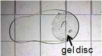 |
(b) | 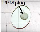 |
(c) | 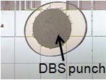 |
Figure 1: Heterogeneous digital microfluidic systems for applications involving liquids and solids. (a) Picture of a droplet containing a proteomic sample interacting with an agarose disc bearing immobilized trypsin. The gel contains 10 μm diameter beads to make it visible. (b) Picture of a droplet of elution buffer (neat acetonitrile) engulfing a PPM plug with C-12 functionality in a solid-phase extraction experiment. (c) Picture of a droplet of extraction solvent (neat methanol) interacting with a 3.2 mm-diameter punch from filter paper bearing dried blood.
Dr. Wheeler is the guest of Prof. Dajana Vuckovic
© Concordia University